We will analyze the structure of the trunk of coniferous trees using a cross section of a pine trunk.
At an early age, the outside of the trunk is covered with periderm, consisting of a multilayer cork, cork cambium (phellogen) and two or three layers of phelloderm. All these parts of the periderm are difficult to distinguish and look like one layer. Later the periderm is replaced by a crust. Under the integumentary tissue there is green parenchyma, or the primary cortex (living storage, and in the presence of chloroplasts, assimilation tissue), made up of loosely arranged round-shaped cells. Among them, large vertical (longitudinal) resin ducts are visible, the channel of which looks like an oval hole lined with living epithelial cells. Later, these resin passages are cut off by a plug and discarded together with the primary bark, so that they are of no importance for the extraction of resin (tapping).
On the inside, the phloem (secondary cortex) adjoins the primary cortex. Pine bast consists of sieve tubes, bast parenchyma and medullary rays (Fig. 13, c). As is known, a downward flow of organic substances from leaves moves through sieve tubes. Sieve tubes look like small (in Fig. 13, h, they are greatly enlarged) oval cells arranged in radial rows, each of which was formed from one cambial cell. Chains of darker rounded cells stretch across the rows of sieve tubes. These are living cells of the storage bast parenchyma, containing starch and other organic substances (fats, proteins), and some cells are crystals of calcium oxalate (drusen), which are waste (see Fig. 14).
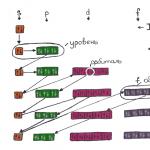
Fig. 13.
a - periderm; 6 - green parenchyma, primary cortex (1 - resin ducts; 2 - parenchyma of the primary cortex); c - secondary bark, bast, phloem (3 - sieve tubes; 4 - bast parenchyma; 5 - medullary ray; 6 - cambium); d - secondary wood, xylem (7 - late (autumn) tracheids; 8 - resin ducts; 9 - early (spring) tracheids; 10 - primary medullary ray; 11 - secondary medullary ray; 12 - primary wood; 13 - core; 14 - growth ring)

Rice. 14. Scheme of the structure of a chloroplast under an electron microscope (a): 1 - stroma; 2 - grains; 3 -- lamellae. Metabolic products in the cell (b):
1-- simple potato starch grains (on the left - concentric, on the right - eccentric); 2 -- complex starch grain of oats; 3 -- crystals; -4 -- crystal intergrowths (druze). Scheme of the gradual formation of vacuoles in a growing cell (c): 1 - cell membrane; 2 - a narrow layer of cytoplasm adjacent to the membrane from the inside; 3 -- vacuole with cell sap; 4 -- core
In the radial direction, medullary rays pass through the bast and continue into the wood. They are chains of living parenchyma cells that are wider in the phloem and narrower in the wood. The medullary rays are the most permanent living part of all trees and shrubs. In woody plants, they represent such an important characteristic element of the anatomical structure that they serve as the main diagnostic feature in the anatomical determination of the wood of various trees and shrubs using a key. The medullary rays arise from the parenchyma cells of the cambium. Water and organic substances move along the cells of the medullary rays in a radial direction (across the growth rings). Some organic substances can be stored in ray cells in the form of reserves of starch and oils. On a transverse section, the medullary rays are visible as lines or stripes crossing the growth rings; on a radial section, as lighter, wide stripes (ribbons) running across the fibers; on a tangential section or on the surface of a trunk with the bark removed, they are visible as darker stripes with pointed ends (strokes) going in the same direction as the wood fibers (Fig. 15).

Rice. 15. Diagram of three sections of the trunk (part of the bark is removed and the tangential surface of the tree is exposed - transverse from above, radial from the side): 1 - bark; 2 -- bast; 3 -- wide core beam; 4 -- annual ring of wood; 5 - cambium; 6 -- core
Under a microscope it can be seen that in a radial section the medullary rays consist of cells elongated along the ray, and therefore across the wood fibers (Fig. 16, A). In a cross section, the rays are rows of elongated narrow cells running in a radial direction. In a tangential section, the rays are cut transversely and look like groups of spindle-shaped small parenchyma cells located between the wood fibers (Fig. 16, B). The rays are strips or ribbons, so they are distinguished on a cross section by length (in the radial direction), width (in the tangential direction) and height (on a radial section in the direction of the fibers). The shorter the ray, the later it was formed from the cambium, and the longer, the earlier. The rays going from the pith through the entire wood to the bark are called primary pith rays. Rays that start not from the core, but in subsequent annual rings of wood, are called secondary. The rays always begin from the cambium, since once formed the ray does not disappear. The medullary rays have two varieties: narrow, visible on a cross section as consisting of a single row of cells, and wide, consisting of several rows. The rays of wood continue into the bast, where they noticeably expand (see Fig. 13).

Rice. 16. Pine wood in a section (A - radial; B - tangential): 1 - early (spring) tracheids; 2 - bordered pores; 3 - late (autumn) tracheids; 4 - core ray (a - - dead tracheid cells with small bordered pores; b - living ray cells with simple pores); 5 - tracheid walls; 6 - bordered pore; 7 - medullary ray; 8 - transverse (horizontal) resin duct in a wide medullary ray; 9 - tracheid cavity; 10 - longitudinal (vertical) resin passage
The cells of the medullary ray are loosely connected. Inside the beam there are always intercellular spaces through which air exchange occurs through the lentils of the bark. This is the only way for the barrel to communicate with the atmosphere.
At the border between the bast and wood there is a secondary educational tissue - the cambium, which is a chain of elongated living cells that form secondary wood inward, and secondary bark (bast) outward. Inside the cambium lies secondary wood, consisting of 90-95% of spring, summer and autumn tracheids, which together form annual rings (layers). The inner part of the annual layer - early or spring wood - contains tracheids that are almost square in cross section with thin shells. Early tracheids have a wide cavity, their radial walls bear bordered pores for communication with each other in a tangential direction, parallel to the direction of the growth rings. This is the main conducting element of the upward current.
The outer part of the annual layer - late wood - has a slightly brownish color and also consists of tracheids. While remaining the same in width, the tracheids are significantly reduced in radius. Their walls are greatly thickened, the cavity is much reduced, there are few bordered pores and they are poorly developed. Late tracheids perform mainly a mechanical function, giving strength to the tree trunk.
The annual rings are intersected transversely by narrow strips that go, like the spokes of a wheel, to the core, which is why they are called medullary rays. In pine wood, especially in the later part of the annual layers, longitudinal, or vertical, resin passages are visible. They have the same structure as in the primary cortex, that is, they consist of three layers: the inner layer of lining, or excretory, cells, forming the epithelium of the resin duct; middle layer of dead cells; the outer layer of living accompanying parenchyma cells. Resin ducts in wood are smaller than in the primary bark, but they function throughout almost the entire life of the tree and are important when extracting resin (tapping) from coniferous trees.
In addition to longitudinal ones, there are transverse, or horizontal, resin passages. They are located in wide core rays and are interconnected with vertical resin passages. In the center of a pine branch lies a core, which looks like a circle with irregular ray processes that turn into primary medullary rays. Small-celled areas of primary wood (xylem), which arose from the procambium, protrude into the space between the rays. In a young pine branch, the pith cells are alive and contain starch.
In a longitudinal (radial) section, pine wood looks different (Fig. 16, A). Early (spring) tracheids have the appearance of long fibers with pointed ends. They bear numerous bordered pores in the form of two concentric circles, located mainly closer to the ends of the tracheids; there are fewer of them in the middle. Late (autumn) tracheids are significantly narrower than spring tracheids, have few bordered pores, which are also much smaller and have only an outer circle, and the inner one is replaced by an oblique slit.
On a radial section, the medullary rays look like ribbons crossing the tracheids in the transverse direction. They consist of two kinds of cells: marginal dead cells, bearing small bordered pores and having thickenings in the form of notches, and middle - living cells, having simple pores in the form of light spots. Dead cells serve to conduct water in a horizontal direction - across the growth rings. Living creatures store reserves of organic substances (carbohydrates, fats, etc.). On a tangential section of pine wood (perpendicular to the radius, Fig. 15, B), medullary rays (cut transversely) are visible in the form of spindle-shaped groups of cells. Transverse, or horizontal, resin passages are visible inside the wide core rays. They, like the vertical ones, are surrounded by an epithelium of thin-walled living parenchyma cells and reinforced by a sheath made of a layer of dead mechanical tracheid cells. Horizontal (transverse) resin ducts, intersecting with vertical (longitudinal) resin ducts, form a dense network called the resin-bearing system of the tree. It permeates all the wood of the trunk, branches and roots.
The connection of vertical and horizontal resin passages allows the resin to flow out of the farthest unopened resin passages due to their connection with the opened ones. This is very important for the extraction of resin by tapping. In the secondary bark (bast) there are only transverse resin passages, which are a continuation of the radial (transverse) passages of wood, since the cambium layer does not interrupt them. Therefore, if you open the resin passage in the bast, resin may flow out of the wood.
Tracheids in a tengental section are dissected along the cavity and therefore early (spring) and late (autumn) ones are very difficult to distinguish and look approximately the same. The bordered pores look like thickenings protruding in two adjacent tracheids. Sometimes a vertical, or longitudinal, resin passage comes into the field of view of the microscope. It has the same structure in radial and tangential sections - it is a hollow canal lined from the inside with epithelial cells.
Lecture. Sheet.
The leaf is one of the most important and specific organs of the plant.
Sheet(Latin folium, Greek phyllon) – lateral, flat ( dorsoventral) an organ that has limited growth and is adapted to perform the functions of photosynthesis, transpiration and gas exchange.
The shape of the leaf helps create maximum photosynthetic surface area.
The role and main functions of the sheet.
1. Main meaning The leaf is that it absorbs solar energy, which is bound in the form of organic compounds and then used not only by plants, but also by all other organisms of the Earth. As K.A. Timiryazev noted, plants play a cosmic role, being intermediaries between space and all other inhabitants of the Earth. And the main role in this mediation belongs to the leaf. Emphasizing this, K.A. Timiryazev wrote that “the whole essence of the plant is expressed in the leaf. A plant is a leaf."
Thus, the leaf is the most important and specific organ of the plant, since it mainly occurs in it photosynthesis process . The primary stem bark also contains chloroplasts, but there are not so many of them and the photosynthetic surface is small.
The leaf is a kind of laboratory for the synthesis of organic substances. 80% of all photosynthesis is provided by leaves.
2. Regulated evaporation of water by leaves (transpiration). Transpiration is not only a physical, but also a physiological process regulated by the leaf.
The main role of transpiration:
1) regulation of the flow of water into the roots and its movement through the vessels;
2) thermoregulation, protecting the plant from overheating.
Plants in forests and deserts, as a rule, evaporate more water than plants in forests, swamps, etc.
3. Gas exchange.
Additional functions:
4. Mechanical function – is expressed in the fact that the soft tissues of the leaf are strengthened by reinforcing mechanical tissues located in the veins, and subsequently the stem of the stem is strengthened and formed due to leaf traces.
5. Storage function not very typical for a leaf. Most often, water can be stored in the leaves (leaf succulents: aloe, agaves, havortia, etc.).
6. Excretory function is provided by various glands located on the leaf and secreting essential oils, water, salts, etc.
7. A leaf may have specific functions during metamorphosis.
General morphological features of the leaf.
1 feature. The main difference between a leaf and a stem and root is that the leaf is not an axial, but a planar organ with large outer surface, which is necessary for efficient photosynthesis, absorption of CO 2 and light, gas exchange and transpiration.
A large outer leaf surface is achieved in two ways: 1) in some plants - by large leaves (banana, palm trees, Victoria Regia), 2) in others - by an increase in the number of leaves (the smaller the leaves, the more of them). A large number of leaves is an adaptive property that helps plants function normally when they are damaged by insects or other animals.
2. The leaf is usually delicate organ, because sunlight must pass through it freely for photosynthesis to proceed normally.
Therefore, the thickness of the leaves depends on the lighting conditions: thin leaves are formed in the shade, thick and denser ones in the light, very thin leaves in the water (since the light is scattered).
3. Unlike the stem and root, the leaf has limited growth. It is typical for a leaf only the primary structure, so that light passes through the sheet well ( and with the secondary structure, opaque and dense conductive and mechanical tissues are formed).
Usually the leaf does not grow for long (in temperate latitudes - 1-2 weeks). The exception is Welwitchia amazing (gymnosperms). Its two leaves grow due to intercalary growth throughout its life (from several hundred to 3 thousand years).
The lifespan of one leaf is short. Deciduous plants have a maximum of one season. There are also evergreen plants whose leaves live for 3-5 years (for spruce - 20 years).
External leaf structure.
Despite the enormous variety of leaves, they usually have 3 (sometimes 2) parts:
1.
 leaf blade
petiole
3. leaf base .
Leaf blade– flattened (more often) part of a leaf.
Leaf petiole– axial, stem-shaped, narrow part of the leaf. The orientation of the sheet relative to the light depends on it. It can change the position of the leaf in space, which is associated with osmotic properties. Leaves with petioles are called petiolate, if the petiole is missing, then the leaf is called sedentary.
Leaf base- part of the leaf with which it is attached to the stem in the area of the node. Through the base, the leaf trails enter the stem.
The base of the leaf can grow:
a) paired outgrowths are formed on the sides of the leaf - stipules. They develop earlier than the leaf blade and protect it for the first time during formation. They are often green and photosynthesize. (Sometimes the stipules are very large and perform the functions of leaf blades, which are reduced or transformed, for example, into tendrils in some legumes). In some plants, the stipules quickly fall off, while in others they remain as long as the leaf lives.
b) the base of the leaf grows and covers the stem, forming vagina . It serves to protect axillary buds and intercalary meristems, and also performs a mechanical function, supporting delicate areas of the stem. The vagina can be in the form of a wide sheath, open on one side (for example, in umbellifers), or in the form of a tube, often closed (in grasses and sedges). In some plants, water accumulates in the vagina, which is then absorbed by the plant (for example, in the tropics - traveler's tree, bromeliads).
The leaves are distinguished by a very wide variety of shapes, dissection of the leaf blade, and leaf edges. (on your own according to the textbook)
By venation there are leaves feathery(oak) (there is one main vein, and secondary veins of the 2nd order extend from it), palmate(maple) (several large veins fan out from the petiole, and from them smaller veins), with arc venation (plantain) and with linear(or parallel) (cereals, sedges). The ancient gymnosperm plant ginkgo and modern ferns have preserved the most primitive type of venation - dichotomous, in which each vein bifurcates, then bifurcates again, etc. This type of venation is found in all extinct and modern spore plants, as well as in a typical or modified form in gymnosperms.
According to the number of leaf blades distinguish leaves simple(with one leaf blade) and complex(with several leaf blades). In compound leaves, the leaf blades are attached to a common petiole - rakhis differently. There are pinnately complex (leaflets sit opposite each other on the rachis) (there are paired and unpaired pinnately compound leaves) and palmately compound leaves (leaflets are attached to the rachis at the apex, like a fan).
Leaf shape is a characteristic feature of the species; however, within one individual, or even one shoot, leaves can vary in shape and form three formations : grassroots, median And riding. Leaves grassroots formation usually in the form of scales, brown or reddish with an undeveloped leaf blade. For example, in lily of the valley they appear first in the spring and perform a protective function. Later, leaves of the middle formation develop on the shoot, which have a normal structure and perform basic functions: photosynthesis, transpiration and gas exchange. On the peduncles, leaves of the upper formation are formed - bracts that protect the buds.
Within the middle formation, the leaves on the shoot can also vary in shape. This phenomenon is called heterophylly (various leaves) . It usually manifests itself in connection with age-related changes or during the life of the plant in different environments and ecological conditions (for example, above-water, underwater and floating leaves of arrowhead differ not only in shape, but also in internal structure; the same in floating pondweed, amphibian knotweed, etc. .; in meadow plants, the upper leaves are narrower, thicker and dissected, receive more light, the lower ones are thinner and wider, less dissected, for example, field bristles).
An even more striking example of leaf differences is anisophilia – leaves vary in shape, size and structure within one shoot node (with opposite or whorled leaf arrangement). Found in plants with creeping or recumbent shoots; the leaves facing the soil are usually scale-like. The aquatic fern Salvinia floating has 3 leaves at a node, two above-water are ordinary, photosynthetic, the third is underwater, dissected and performs a suction function.
Anatomy of a leaf.
The anatomical structure of the leaf is a hereditarily fixed trait.
Leaf ontogeny.
The leaf is laid at the apex of the shoot. Below the growth cone, tubercles of rudimentary leaves appear - leaf primordia . Each primordium appears after a certain strictly constant period of time - plastochron (“plastos” - shaped, “chronos” - time).
For example, oak has a plastochron of 2.8 days, linden has 5 days, and spruce has 4.3 hours. As a rule, the smaller the leaves, the shorter the plastochron. Thanks to the plastochron, the leaves on the plant are arranged in a strictly defined order.
Leaf growth is different from stem and root growth. 
1) A new leaf primordium is formed inside the bud in the form of a meristematic tubercle, which initially The apex grows for a very short time (apical growth). Then the leaf primordium differentiates into upper and lower parts, which grow unequally.
2) The leaf actively grows at the base (procambium is formed). The base of the leaf is formed.
3) The area of the central vein grows intercalated in length, thickens and acquires a cylindrical shape (the leaf axis is formed).
4) On the sides of the leaf axis, a leaf blade begins to form due to the marginal (marginal) lat. Margo - edge) diffuse growth. Marginal meristems are laid in the form of ridges on the sides of the central vein and form the plane of the leaf. Unevenness of marginal growth leads to the formation of blades with uneven edges, lobes, dissected, etc. Stipules, as a rule, form before the leaf blade (as outgrowths of the leaf base) and protect it from damage.
5) At the last stage, the petiole grows. It appears after the leaf blade finishes growing. The petiole grows in length due to intercalary growth (intercalary growth) and in a certain way orients the leaf in relation to the light.
Anatomy of a typical leaf.
In the anatomy of a leaf, the connection between the anatomical structure and the functions performed is clearly visible: photosynthesis, transpiration and gas exchange.
Anatomical differences between a leaf and a stem and root.
1) The sheet is dominated by parenchymal tissue, but not storage parenchyma, but highly specialized assimilation parenchyma, the cells of which contain chloroplasts.
2) There are few conductive and mechanical tissues; they form veins leaf.
3) Many intercellular spaces (related to the gas exchange function).
4) Various excretory tissues may be developed.
The leaf has only a primary structure!
1. The outside of the leaf is covered with primary integumentary tissue - epidermis. On the leaf, the epidermis has its most typical structure. A thick layer of cuticle protects against excessive evaporation of water. (in plants in humid habitats the cuticle is thin or absent), as well as trichomes of various structures. There are a lot of stomata. Most of the stomata are on the underside of the leaf. This is explained by the fact that 1) with open stomata on the upper side of the leaf, a lot of water would be lost; 2) the main source of CO 2 is the soil, where organic matter decomposes and carbon dioxide enters the atmosphere. It is heavier than air and usually accumulates in its lower layers. Since carbon dioxide rises from the bottom up, the location of the stomata on the lower side facilitates its rapid entry into the leaf along the shortest path.
Sometimes stomata may be evenly distributed on both sides of the sheet if the leaves are located edge-on to the sun (in a number of savannah and desert plants, in eucalyptus - tree that doesn't provide shade).
Only on the top side of the sheet Stomata are located in aquatic plants with leaves floating on the surface of the water. Leaves that are completely submerged in water do not have stomata.
Number of stomata– on average per 1 mm 2 - 250 stomata.
In some plants (usually growing in arid climates), under the epidermis there may be a hypodermis (colorless), the cells of which perform a water-storing function, and less often a mechanical function (for example, in conifers).
2. Leaf mesophyll – highly specialized assimilative leaf tissue.
In most flowering plants, mesophyll cells are not uniform in shape. They have 2 types of mesophyll: 1) columnar (palisade), adjacent to the upper side of the leaf; 2) spongy (loose), adjacent to the underside of the leaf.
Columnar mesophyll consists of closed cells elongated perpendicular to the surface of the leaf. This tissue receives more light and the maximum number of chloroplasts is concentrated in it. 80% of photosynthesis occurs here.
The shape of the cells is not random:
1) Thanks to this shape, chloroplasts are protected from very bright sunlight. With a sharp increase in light intensity, chloroplasts move from short walls to long walls, perpendicular to the surface of the leaf.
2) It is necessary that the organic substances formed in the leaf cells are quickly removed. With this form of cells, the outflow of assimilated substances occurs quite quickly.
Spongy mesophyll- loose tissue, with a large number of intercellular spaces. The cells are usually round and have much fewer chloroplasts. Less light reaches them and only 20% of photosynthesis occurs in spongy tissue cells. Nevertheless, The value of this fabric is very great, precisely thanks to the developed system of intercellular spaces, which contribute to 1) transpiration, because water vapor is released into the intercellular spaces from surrounding cells; 2) gas exchange(CO 2 entering through the stomata through the intercellular spaces quickly spreads throughout the leaf, the released oxygen spreads through the intercellular spaces and exits through the stomata ( when breathing in reverse)) normal photosynthesis.
If the leaf faces the light with its edge, then columnar tissue develops on both sides of the leaf.
The distribution of spongy and columnar tissue depends on lighting. The greater the illumination, the more developed the columnar tissue. With shading, spongy tissue develops more strongly and transpiration will occur more strongly.
Therefore, the upper (outer) leaves ( light) and lower (located deep in the tree crown) ( shadow) have different ratios of columnar and spongy tissue.
Light the leaves are small and thicker, with a powerful cuticle, well-developed columnar tissue.
Shadow the leaves are thinner and larger, the columnar tissue is poorly developed, often 1 layer, the cells have the shape of funnels, directed with the wide side towards the surface of the leaf, spongy tissue predominates.
In most monocots and some dicotyledons and conifers, the mesophyll is uniform and not differentiated into columnar and spongy. This mesophyll is called isopalisade.
3. Conductive and mechanical fabrics - form the veins of the leaf.
Veins are collateral, closed vascular-fibrous bundles. Phloem in the bundle facing bottom side of the sheet, and xylem - To top. Bottom and top they are reinforced with sclerenchyma fibers. The veins of the casts branch, and the smaller veins have a simpler structure. They have no mechanical tissues (only sieve tubes and vessels). In some plants, thin veins consist only of tracheids, which are in direct contact with the leaf tissues. In addition to transporting water, substances - assimilates - move through these tracheids. In other plants, thin veins may consist only of sieve-like elements, tubes have unclear “strainers” or none at all, companion cells may disappear, and sometimes become larger. At the terminal sections, such veins are represented only by mother phloem cells, not differentiated into sieve tubes and companion cells. Modern research has revealed several types of structure of small veins (see textbook.)
Many plants have a lining of parenchyma cells around the veins. These cells are elongated along the veins and do not contain chloroplasts. Through them, the products of photosynthesis enter the veins.
Sometimes the mechanical tissue of the veins is not enough, and then additional mechanical fabrics. In large veins, collenchyma is added above and below (it is often present in leaf petioles), and sometimes additional sclerenchyma develops. The mesophyll can be strengthened by sclereids - idioblasts scattered between assimilation tissues. Some plants develop a lot of bast fibers in the leaf blade (agave, palm trees, banana, traveler tree).
4. Excretory tissues - glands with essential oils, containers of resins, lactic acids, hydathodes, etc.
Anatomical structure of a conifer leaf.
Conifers arose in the late Carboniferous (about 290 million years ago), when the climate on the planet began to dry out. The leaves of modern conifers have many features indicating their drought resistance, i.e. have xeromorphic features. This may be due to the fact that most representatives of this class were finally formed during the dry and relatively cool Permian period (286 - 248 million years ago). At that time, the gradual increase in aridity probably favored these types of structural adaptations.
The leaves of conifers are needle-shaped (pine, spruce, fir, larch) or scale-like (thuja, cypress), usually evergreen ( excl. Larch is a secondary adaptation to very cold climates), adapted to economical transpiration of water and to endure drought, including winter, when at low temperatures the roots cannot absorb water.
1) The outer surface of the needles is very small (small evaporation area).
2) The epidermis consists of thick-walled cells with a powerful cuticle (protection against evaporation).
3) Submerged stomata. The guard cells partially become lignified, and the canal is filled with resins or wax (a sharp decrease in transpiration).
4) Under the epidermis in a ring there is a special hypodermal tissue consisting of lignified fibers, which reduces evaporation and increases mechanical strength.
5) The main difference from angiosperms: there is no differentiation into columnar and spongy mesophyll, all cells are homogeneous, form folded mesophyll. This is adaptive compensation for the small outer surface. In mesophyll cells, the membrane forms internal folds, which ensures a sharp increase in the wall layer of the cytoplasm and the inner surface.
In cells, due to an increase in their internal surface, the number of chloroplasts increases, and with a small external surface of the needles, photosynthesis processes proceed as intensively as in ordinary leaves of flowering plants.
6) The double vascular-fibrous bundle is surrounded by endoderm, which regulates the transport of substances. When entering the stem, the double bundle merges into one, forming a single leaf trail.
7) Conducting bundles are surrounded by transfusion tissue, which consists of: a) ray tracheids (water transport), b) living parenchyma cells (transport of organic substances-assimilates).
8) There are resin passages located between the mesophyll cells.
Leaf metamorphosis.
A sheet can perform functions that are not typical for it:
1) The role of the root (salvinia - in the form of thin threads in water - similar organs)
2) Leaves often turn into spines (in plants of deserts, steppes, savannas). The mesophyll of the leaf is reduced, and usually only the central vein remains, which is additionally reinforced by sclereids (cacti, barberry, etc.).
What are spines an adaptation to: a) reducing the evaporating surface under conditions of water deficiency; b) protection from being eaten by animals.
Using an electron microscope, it was possible to determine the role of cactus spines. These are microscopic pumps that draw in air and condense water.
3) Transformation of leaves into tendrils (legumes, pumpkin) - serve to support the stem and attach it.
4) Sometimes the petiole may change. It flattens, turns green and performs the function of photosynthesis. In this case, the leaf blade is often reduced. Characteristic of plants in arid areas (for example, Australian phyllodes acacias)
5) Storage leaves - most often water (aloe, agaves, etc.).
6) In insectivorous plants, leaves turn into trapping devices. They usually have digestive glands that secrete digestive juice.
These plants were first studied by Charles Darwin. He explained the appearance of these plants. They live where the soil is low in nitrogen, phosphorus and other minerals (for example, in peat bogs, tropical rainforests, standing ponds). They obtain the substances they need by digesting insects and sometimes other small animals.
About 500 species of insectivorous plants (predatory plants) are known. They are found from the Arctic to the tropics. There are 3 groups of insectivorous plants, differing in the types of traps. These are: 1) traps(sarracenia, nepenthes); 2) Velcro(sundew, rosewort, etc.); 3) traps(Venus flytrap, pemphigus).
1. traps . U sarracenia(North America) trapping leaves resemble pitcher flowers. They are brightly colored and have a landing area for insects on the outside, and nectaries at the entrance to the pitcher. Sharp hairs pointing downwards are also located here, which allow the victim to easily slide down, but do not allow them to rise up. The jug is 2/3 filled with liquid. The walls of the jug have digestive glands that secrete digestive juice. More primitive dovushki are filled with rainwater, the insects that get there first decompose and then are absorbed by the plant.
Nepenthes(trop. Asia) - a very narrowly specialized liana. The leaf petiole consists of 3 parts: the phyllode, the petiole itself and the brightly colored pitcher; the pitcher is covered by a colored leaf blade. The jug can hold up to 1 liter of liquid containing digestive juice. There are nectaries to attract insects, the walls of the jug are covered with wax and hairs pointing downwards. Once trapped, the insect is digested in 5-8 hours.
2. Velcro. Sundew leaves are covered with glandular hairs that secrete a sticky secretion. There are also digestive glands. Droplets of liquid glisten in the sun like drops of dew, attracting prey. There is no nectar, no smell. Insects land on the leaf and stick to it, the leaf curls and secretes digestive juice. Digested food is absorbed. After a few days, the leaf unfolds.
3. traps . The most complex ones are found in the Venus flytrap (North America). The leaf is divided into two parts, the upper one is a trap, on it there are sensitive and glandular hairs. As soon as insects touch the hairs, the leaf instantly collapses (turgor!).
A complex trap for bladderwort. (Kr. Book St. region - 3 types). On the thin, dissected leaves floating at the surface of the water there are numerous trapping bubbles (up to 2 mm in diameter). The trapping vesicle has a round hole with a valve and sensitive hairs. There is negative pressure in the cavity of the bubble, since all the liquid is pumped out of it. Small crustaceans (daphnia, cyclops), ciliates, swimming by, touch the sensitive hairs, the valve instantly opens and the prey is sucked into the bubble along with water. The valve closes.
Leaf structure (needles) of Scots pine (Pinus sylvestris L.)
Scots pine has hard, needle-like leaves (needles) arranged in pairs on short shoots.
The needles are fixed with alcohol, which partially dissolves the resin they contain. To make cross-sections easier, pairs of needles are sandwiched between pieces of elderberry core or stuck into the core. Thin sections are treated with a solution of phloroglucinol and hydrochloric acid.
The cross section of the leaf has a semicircular outline (Fig. 89). On the outside is the epidermis with a thick cuticle. Epidermal cells are almost square. The outer, lateral and inner walls of the cells are greatly thickened; in the oldest leaves they often become lignified. Narrow slit-like pore channels extend from the small round internal cavity to the corners of the cell. Under the epidermis is the hypodermis, consisting of one, and in the corners - two or three layers of fibers with thickened lignified walls.
Stomata are located over the entire surface of the leaf. Their guard cells are located at the level of the hypodermis, under the parostomatal cells. The peristomatal cells are very large, with very thickened outer walls. The walls of guard and parastomatal cells in thickened areas become lignified. The stomatal fissure leads into the substomatal air cavity, surrounded by mesophyll cells.
The mesophyll is homogeneous and folded. Folds arise due to the ingrowth of the inner layers of the membrane into the cell cavity, which at the same time acquires a lobed outline. Due to the folds, the surface of the wall layer of cytoplasm containing chloroplasts increases. Mesophyll cells are tightly connected, the intercellular spaces between them are very small.
In the mesophyll, directly under the hypodermis or somewhat deeper, schizogenic resin canals are located. They run along the leaf and end blindly near its tip. On the outside, the resin channel is lined with thick-walled, non-lignified fibers. Inside, it is lined with thin-walled living epithelial cells that secrete resin.
The conducting system is represented by two collateral closed bundles located in the center of the needles at an angle to one another. The xylem, consisting of tracheids with narrow cavities, faces the flat side of the leaf, the phloem faces the convex side. Thus, the flat side of the needles represents the morphologically upper side, and the convex side represents the morphologically lower side of the leaf.
Below, between the bundles, there is a strand of fibers with thick, slightly lignified walls. Conducting bundles and adjacent mechanical elements are surrounded by transfusion tissue, consisting of two types of cells. Near the xylem, the cells are somewhat elongated, they have no contents, their lignified walls have bordered pores. These cells are called transfusion tracheids. The remaining cells are living, parenchymal, thin-walled. They contain resinous substances and often contain starch grains. Transfusion tissue appears to be involved in the movement of substances between vascular bundles and the mesophyll.
The vascular bundles, together with the surrounding transfusion tissue, are separated from the mesophyll by the endoderm, which is a single-row layer of parenchyma cells with Caspary spots on the radial walls.
Metamorphoses of vegetative organs root leaf shoot
Root
Microscopic structure of the root
. In a longitudinal section of a young growing root, you can see: the division zone, the growth zone, the absorption zone and the conduction zone. The apex of the root, where the growth cone is located, is covered by a root cap.
Root function and root systems
. The main functions of the root: anchoring the plant in the soil, actively absorbing water and minerals from it, synthesizing important organic substances, and storing substances.
The totality of all the roots of one plant forms the root system.
There are two types of root systems - taproot, in which the main root is clearly visible, and fibrous, consisting of adventitious roots.
Root modifications. In the modified roots, reserve nutrients accumulate - starch, various sugars and other substances. The thickened main roots of carrots, beets, and turnips are called root vegetables. The adventitious roots also thicken, as, for example, in the dahlia. These are called root tubers.
The escape
During the evolution of plants, during their transition to terrestrial existence, a vegetative organ was formed - a shoot, which performs the functions of photosynthesis and the formation of reproductive structures (sporangia, cones, flowers, etc.). A shoot is a stem bearing leaves and buds.
Development of escape from the buds
. The above-ground part of the plant usually consists of a system of branching shoots. The stem is the axis of the shoot; it connects the roots and leaves. Shoots can be annual or perennial. The stems of annual plants usually do not become lignified, but those of perennial plants do. The shoot develops from the bud of the seed embryo. A bud is a rudimentary shoot consisting of a shortened stem with rudimentary leaves. It is covered with scales that fit tightly together, which protect it from adverse influences.
There are vegetative and generative (floral) buds. Flowers form from flower buds. Vegetative - leaves and shoots. The apical bud is the tip of the stem. The very tip of the stem is called the growth cone. The main shoot grows from the apical bud, and lateral shoots from the lateral buds.
Plants can form buds on any part of the stem, on the roots and even on the leaves.
Stem branching. In the process of evolution of higher plants, the following main methods of branching were developed: dichotomous, or forked, monopodial, sympodial.
Dichotomous branching. Two shoots extend from the top, each of which, in turn, gives rise to two more shoots, etc. (moss mosses, some fern-like ones).
Monopodial branching. The main axis, the monopodium, has seemingly unlimited apical growth. Lateral axes of the second order extend from the monopodium, giving rise to axes of the third order, etc. (many gymnosperms).
Sympodial branching. One or several side shoots formed on the main shoot quickly outstrip its growth (pear, linden, shrubs).
Stem shapes. The forms of shoots are varied: erect, creeping, curly, climbing. There are herbaceous and woody stems that form the corresponding life forms of plants (annual and perennial herbs, trees and shrubs).
Stem modifications. The stem can serve as a nutrient reserve. At the same time, it is modified, forming rhizomes, tubers, bulbs, etc. A rhizome is a highly modified underground shoot that develops scale-like leaves and buds (in this way it differs from the root). Adventitious roots form on it. The bulb consists of a strongly shortened stem - the bottom, from which a bunch of adventitious roots extend downwards, and the shortened stem is surrounded by modified thick leaves, which form the pulp of the bulb. The rhizome, tuber and bulb serve as organs of vegetative propagation.
Sheet
The leaf implements three important functions: photosynthesis, water evaporation and gas exchange.
The leaf consists of: leaf blade and petiole. Leaves that do not have a petiole are called sessile.
Based on the shape of the leaf blade, leaves are divided into round, lanceolate, heart-shaped, kidney-shaped, arrow-shaped, etc.
Leaves are divided into simple and compound. Simple sheet consists of a petiole and a leaf blade; compound leaves have several leaf blades located on one petiole. Simple leaves can be entire or lobed. Many trees (birch, linden) have whole leaves. In lobed leaves, the blade has cuts that divide it into lobes (maple, oak). Compound leaves are palmate, trifolate and pinnate. In the latter, leaf blades are attached along the entire length of the petiole. They come in two types: pari-pinnate and odd-pinnate. The pinnate ones end in a pair of leaf blades (peas); imparipinnate - one leaf (rowan, ash, raspberry).
Simple and compound leaves are arranged on the stems in a specific order. The regular arrangement is characterized by the fact that the leaves sit on the stem one at a time, alternating with each other (birch, apple tree, rose). With an opposite arrangement, the leaves are placed two opposite each other; with a whorled arrangement, they are attached to the stem in bunches - whorls.
Leaf structure. The leaf blade is covered with skin. On the underside of the leaf there are stomatal cells that limit the stomata. Under the skin there are leaf pulp cells - columnar and spongy tissue. The leaf tissue is also represented by a system of conducting bundles - veins. Through them, water, mineral elements and substances formed in the roots are delivered to the leaves. Substances formed during the process of photosynthesis flow from the leaves to the stem to the buds and roots. There are reticulate (most often found in dicotyledons), parallel (in monocotyledonous grasses, sedges) and arcuate (for example, in lily of the valley) venation.
Evaporation of water by leaves
. Evaporation promotes the movement of water and dissolved substances from the roots to the leaves. The intensity of evaporation is regulated by stomata. Light promotes the opening of stomata; in the dark they are closed. The stomata also close in the middle of the day, in extreme heat.
Leaf modifications
. In the process of evolution, leaves acquired additional functions, and therefore their appearance changed. For example, cactus and barberry leaves have turned into spines. In peas, the leaves have changed into tendrils, through which the plant is attached to the support. In scaly leaves of a bulb (for example, onions), thin scales play a protective role, and succulent scales, rich in nutrients, serve as storage organs.
Conifers are the oldest existing plants on our planet. Their age is estimated at hundreds of millions of years. Evolution has had virtually no effect on the anatomical structure of needles and cones. When comparing the leaves of conifers, which are popularly called needles, with the leaves of flowering plants, one can notice that while the needles are relatively uniform, they have different shapes, sizes, colors, and in some species they do not look at all like ordinary needles.
The needles look like narrow needle-like leaves. It is characterized by the presence of a dense skin, which is covered with a waxy substance. This is necessary to reduce the evaporation of moisture by gymnosperms. For example, spruce needles are tetrahedral, but often the edges are almost invisible, and the needles look flattened.
Drawing. Cross section of Scots pine needles
If you cut a needle, it has the shape of an irregular diamond, with the flattest angle directed downward. This is where the midrib of the leaf is located. Along the other edges of the needle, white stripes are visible, formed by stomata - respiratory openings through which plant respiration occurs. Stomata also serve to evaporate moisture, which the tree absorbs from the soil even in severe cold. This explains the fact that spruce, like other conifers, cannot be replanted in the autumn, since the roots cannot take root firmly, and water practically does not rise up the stem to the needles, although respiration occurs in the same mode.

An important difference between conifers and deciduous trees is that their petiole is firmly connected to the branch and remains on it, even after the needle dies. The needles fall off after 6-7 years. They are well protected from the effects of adverse environmental factors by a thick layer of waxy coating - the cuticle. Moreover, in many species the coating is so thick that the needles acquire a blue tint.
Conifers do not have true fruits and flowers. They belong to the division of gymnosperms.
Their seeds are attached directly to the seed scales, and those that are collected into female cones are equipped with special wings. Leaving the cone, they glide on their wings, resembling small helicopters when rotating. This helps them move away from the mother plant.
The appearance of conifer cones is varied and specific. They may differ in length, shape, placement in space, color, structure and shape of sporophylls, method of seed dispersal, etc. But the fundamental structure of the cones is the same. All cones at the base have an axis, which is separated from the vegetative part of the tree and is a short shoot with spore-bearing leaves located on it - sporophylls.

There are female and male cones. The vast majority of conifers are monoecious. They have both female and male cones developing on the same plant. In most cases, male cones are concentrated in groups in the axils of the leaves, sometimes on the tops of the side shoots. Female cones are distinguished by a compact arrangement, occasionally they are located singly.
Related materials:
Goal of the work: study the anatomical structure of Scots pine needles and the rhizomes of common bracken fern.
Required materials and equipment: microscopes, permanent preparations, herbarium specimens.
Tasks:
1. Consider a cross section of a leaf (needles) of Scots pine ( Pinus sylvestris L.) at low and high magnification microscope. Draw a diagram of the structure of a cross section of a needle at low magnification, noting the hypodermis, resin canals, mesophyll, endoderm, vascular bundles, and transfusion tissue. At high magnification, sketch a section of the cross section, on which to designate the mesophyll, endoderm, hypodermis, epidermis, and resin canal.
2. Examine a cross section of a bracken rhizome ( Pteridium aquilinum(L.) Kuhn.) at low and high magnification microscope. Draw a cross-section diagram at low magnification, noting vascular bundles, areas of mechanical tissue, and the zone of outer and inner cortex. At high magnification, sketch the vascular bundle, showing the endoderm, pericycle, phloem and xylem.
Leaf structure of Scots pine
 For pine needles ( rice. 70), like the perennial leaves of other conifers, are characterized by a xeromorphic structure, the development of which is caused by sharp changes in temperature throughout the year and the need to reduce evaporation in the winter, when the water supply is insufficient.
The leaves are needle-shaped and arranged in groups of 2 on short shoots.
The cross-section of the leaf is semicircular, its upper side is flat, and its lower side is convex.
On the outside is the epidermis, consisting of almost square cells covered with a layer of cuticle. The walls of the epidermal cells are very thickened and lignified. As a result of thickening, only a small cavity remains inside the cells, from which narrow pore channels extend diagonally to the corners.
Under the epidermis is a one- or two-layer hypodermis, consisting of flattened cells with evenly thickened walls. At the level of hypodermal cells, stomatal guard cells with lignified walls are visible.
Under the hypodermis layer, resin canals are visible, lined with thin-walled epithelial cells and surrounded by a lining of thick-walled cells.
In the center of the leaf there is a conducting system, which consists of 2 collateral bundles, connected by mechanical tissue and surrounded by transfusion tissue (serves to move water and solutions of organic substances from the bundles to the mesophyll). Outside the conducting system is a single-row endoderm.
Mesophyll consists of cells of unusual shape: they have numerous folds that appear as a result of the invagination of the membrane into the cell, which serves to increase its surface.
The structure of the bracken rhizome
The long horizontal rhizome of bracken is located at a depth of 20-40 cm underground. The roots of the bracken are black and extend downwards from the rhizome. The rhizome itself has a polycyclic structure ( rice. 71).
In a cross section, two large oval vascular bundles are clearly visible in the center. Around them are two half-rings of mechanical fabric. Behind it are small round bunches, among which one large oval one is usually visible.
The bundles are immersed in parenchymal tissue, where the inner and outer cortex are clearly visible. The outside of the stem is covered with epidermis.
Numerous small tufts are visible in the bark, also extending into the leaves. Bracken bundles are closed; they are delimited from the bark by endodermis with passage cells. Next is the pericycle, then the phloem, in which sieve tubes and bast parenchyma are visible. In the center of each bundle is xylem.


Questions about the material covered:
1. Describe the features of the anatomical structure of leaves.
2. Describe the features of the anatomical structure of rhizomes.
3. Describe the features of the anatomical structure of plants from various ecological groups (hydrophytes, hygrophytes and xerophytes; heliophytes - sciophytes; succulents - sclerophytes).