Assistant of the Department of Pediatrics, Ph.D.
Zaitseva L.Yu.

POLIO
HISTORY OF THE ISSUE
Poliomyelitis is a disease known since ancient times. In the temple of the fertility goddess Astarte in Memphis, they found something made in 1580 BC. image of the priest Ruma with a lower limb lesion typical of polio

POLIO
HISTORY OF THE ISSUE
In the 19th century, it was finally established that polio epidemics were possible.
In 1836, pathologist Charles Bell identified a number of cases of infantile paralysis in an isolated community on the island of St. Helena
In 1840, the German orthopedist Jacob von Heine for the first time described in detail a disease that was rare at that time - infantile paralysis

POLIO
HISTORY OF THE ISSUE
In 1900, Mitchell discovered bone changes characteristic of polio in a well-preserved mummy.
In Greenland, during excavations, skeletons dating back to 500–600 AD were found.
BC. and having the same bone changes
Hippocrates (460–377 BC) described an outbreak of polio on the island of Pharos

POLIO
HISTORY OF THE ISSUE
In the mid-twentieth century, the incidence of polio increased sharply in the USA, Canada, South America, Scandinavia, Central Europe, and Asia.
In the United States, the epidemic almost did not stop in the period 1942–1953, with paralysis affecting up to 20 thousand people every year. In 1956, more than 300 thousand disabled people were registered as having suffered a paralytic form of the disease.
Almost at that time, the entire population of the country was infected, because... For each disease that causes paralysis, there are 100 to 1000 cases of asymptomatic infection

POLIO
HISTORY OF THE ISSUE
1947 Jonas Edward Salk took up the problem of obtaining serum against polio
He was able to prove that there are three serotypes of polioviruses
After three doses of the vaccine, antibodies to all serotypes of the virus are detected in the blood
Jonas Edward Salk (1914–1995)

POLIO
HISTORY OF THE ISSUE
An important step in improving the polio vaccine was made by A.B. Sabin
In 1939, he proved that poliovirus enters the human body through the digestive tract
He was convinced that a live vaccine, taken orally, would produce longer-lasting and more reliable immunity.
Albert Bruce Sabin (1906–1993)

POLIO
HISTORY OF THE ISSUE
US President F.D. Roosevelt suffered as an adult
polio, after which he could only move in a wheelchair
The Roosevelt family created a large fund to support polio research
Salk received $1 million from this fund to carry out his work, which allowed him to complete his research.

POLIO
HISTORY OF THE ISSUE
The live vaccine received final recognition after in the USSR, as a result of the work of our wonderful scientists M.P. Chumakov and A.A. Smorodintsev were held
Slide 2
1. Features of the disease………3-6 2. Symptoms………………………….…7-10 3. Treatment…………………………..…11-12 4. Prevention …………………….13-17 5. Literature ……………………..……..18
Slide 3
Features of the disease
Poliomyelitis - from the Greek words polios - gray, myelos - spinal cord. Poliomyelitis (Heine-Kozhevnikov-Medina disease) is an acute infectious disease of viral etiology, characterized by damage to the gray matter of the spinal cord with the development of flaccid paresis and paralysis. There are three known antigenic types of poliomyelitis, designated by numbers I, II, III. I and III types of poliomyelitis are pathogenic for humans and monkeys, and type II is also for some rodents. A.Ya.Kozhevnikov (1836 -1902)
Slide 4
Facts from the history of the disease
There are known cases of the disease in Ancient Egypt and Ancient Greece - several centuries BC. In 1840 and 1860 surgeon Heine described this disease and proposed to call it infantile spinal paralysis. At the end of the 19th century, A. Ya. Kozhevnikov and Medin gave additional descriptions of various forms of polio and pointed out the infectious nature of the disease. The virus, the causative agent of polio, was discovered by Viennese scientists in 1909 by K. Landsteiner and E. Poper. In 1954-1955 Vaccinations began with the inactivated (killed) J. Salk vaccine, and 4-5 years later with a more effective drug - the live A. Seibin vaccine, which includes all 3 antigenic types of the virus. Soviet scientists A. A. Smorodintsev and M. P. Chumakov took an active part in the creation and improvement of the live vaccine.
Slide 5
Routes of infection
airborne transmission mechanism fecal-oral transmission mechanism
Slide 6
Features of the course of the disease
The entry point for poliovirus is the mucous membranes of the oral cavity. During the incubation period (from 2 to 35 days, usually 5-14 days), the virus multiplies in the intestinal epithelium, lymphatic formations of the pharynx and intestines. Next, the pathogen enters the brain tissue, revealing tropism for motor neurons. The virus can enter the central nervous system along the axons of nerves. Poliovirus causes the death of affected cells. When a significant part of the nerve cells dies, paralysis occurs.
Slide 7
Forms of polio
Non-paralytic: Abortive form Meningeal form Paralytic: Paralytic form Spinal form Pontine form Encephalitic form
Slide 8
Symptoms of the non-paralytic variant of polio
Abortive form: Increase in temperature to about 38°C; Weakness; General malaise; Mild headaches; Lethargy; Stomach ache; Runny nose; Cough; Vomit. Meningeal form: Symptoms similar to the abortive form Sudden deterioration in the general condition of the patient; Severe headaches; Pain in the back, limbs (mainly in the legs); Stiffness of the neck and neck muscles
Slide 9
Symptoms of the paralytic form of polio: Convulsive contractions in the muscles with characteristic pain Muscle weakness Sweating The appearance of paresthesia (sensitivity disorder with tingling, numbness and goosebumps) Atrophy, deformation, curvature of the spine and shortening of the limbs
Slide 10
Depending on the location of the lesion, the following types of paralytic forms of poliomyelitis are distinguished:
Slide 11
Treatment of polio
The main treatment for polio is complete rest and bed rest. Such conditions can significantly reduce osteoarticular complications in case of paralysis. Sometimes patients are recommended support corsets, bandages, etc. In many cases, it is advisable to prescribe physical therapy: a variety of muscle warming, special physical therapy. Medicines that are usually used are drugs that have anti-inflammatory and analgesic effects, as well as tranquilizers.
Slide 12
The therapy carried out should be aimed at restoring and strengthening weakened muscles.
Correct position of a polio patient. Rice. 3. On the back. Rice. 4. On the stomach. Rice. 5. Drainage pose. Rice. 6-13. Therapeutic exercise in the treatment of the consequences of polio. Rice. 8. Initial position of the patient, facilitating movements in the knee joint. Rice. 7. The instructor supports the patient’s limb with straps to facilitate movements in the knee joint. Rice. 8. Performing movements in the hip joint while supporting the patient using straps. Rice. 9. Abduction of the patient’s lower limb along the vinyl plastic surface, facilitated by the traction of the load. Rice. 10. Facilitating hip flexion by balancing the limb with a counterweight. Rice. 11. Exercise on a gymnastic wall for the muscles of the upper belt. Rice. 12. Using a standing aid. Rice. 13. Use of a device for learning to walk.
Slide 13
Prevention of polio
Specific prevention is vaccination against polio. There are 2 types of polio vaccines: 1. Live Sebin vaccine (OPV - contains live attenuated viruses) 2. Inactivated (IPV - contains polioviruses of all three serotypes killed by formaldehyde) Salka.
Slide 14
Precautionary measures after OPV vaccination: do not kiss the child on the lips and wash your hands after washing the baby.
Contraindications for OPV vaccination: - children with congenital immunodeficiency or HIV - the presence of pregnant women around the child; - pregnancy or planning it; - breastfeeding; - an unusual reaction to a previous vaccination; - acute infectious diseases (vaccination after recovery).
Slide 15
IPV vaccine
IPV vaccination is carried out: - children (weak, with a pregnant mother and/or intestinal disorders) - adults (medical workers who have close contact with patients, travel to endemic areas, unvaccinated people). IPV is administered subcutaneously or intramuscularly: - children: primary vaccination at 2, then at 4 months, then revaccination at 6 - 18 months and at 4 - 6 years; - adults: first vaccination (0.5 ml), repeat after 4 - 8 weeks and a third dose after 6 to 12 months.
Slide 16
Side effects of IPV vaccination: Possible side effects that do not require emergency medical intervention: - nervousness, - fever up to 38.5°C, - swelling, - pain at the injection site, - nausea, single vomiting or diarrhea. Consult a doctor immediately case: - adynamic and lethargic child; - difficulty breathing, shortness of breath; - temperature above 39 degrees; - convulsions; - urticaria, itching; - drowsiness; - swelling of the face, eyes; - difficulty swallowing. After vaccination with IPV, walking and bathing the child is not are prohibited.
Slide 17
At the source of the disease, sanitary and hygienic measures are carried out - disinfection of dishes, clothing, and all items that could be contaminated. Contact children are quarantined for up to 15–20 days.
The last cases of polio in Russia were registered in 1996 in Chechnya due to the lack of vaccinations among the population.
Slide 18
Literature
Badalyan L.A. “Neuropathy” Drozdov S.G. Poliomyelitis and its prevention, M. - 1967. Voroshilova M.K. Immunology, epidemiology and prevention of polio and similar diseases. M., 1966. Ganzburg S.E. Poliomyelitis. In the book: Guide to Pediatrics. M. 1965, vol. VIII, ch. VII, p. 105. Conburn V. Ch., Drozdov S. G. Further observations of the global spread of polio. Question Virol., 1968, 5, 531. Futer D. S. Acute poliomyelitis. In the book: Multi-volume guide to neurology. M., 1962, vol. 3, v. 1, p. 137. Footer D. S. Diseases of the nervous system in children. M., 1965. Zucker M. B. Infectious diseases of the nervous system in children. M., 1963, ch. II and III, p. 67 and 198. Zucker M. B., Leshchinskaya E. V. On the clinic and diagnosis of non-paralytic polio. Pediatrics, 1957, 3, 13. Chumakov M.P., Prisman I.M., Zatsepiv T.O. Poliomyelitis - infantile spinal paralysis, M., 1953. Chumakov M.P. Poliomyelitis. Etiology. BME, vol. 25, 162, p. 756.
View all slides
POLIOMYELITIS1980
year
–
350
000
cases
paralytic polio in the world;
number of countries – 250;
After the creation of the Global Initiative
to eradicate polio – reduction
incidence by 99%, the number of endemic
countries decreased to 3 (Afghanistan,
Pakistan, Nigeria)
2012 – 223 cases of polio
2013 – 403 cases of polio
3
Poliomyelitis virus, a genus of enteroviruses,
RNA-containing.
Resistant to ether and alcohol. Up to 3-4 months
persists in feces, wastewater,
on vegetables and milk. Sensitive to
high T (quickly dies at
boiling), ultraviolet radiation and disinfectants. Epidemiology.
The source of infection is the patient or the carrier. The virus is isolated from
nasopharynx up to 5 days from the onset of the disease, and from feces - from
several weeks to 3-4 months. Particular importance in distribution
have virus carriers. For 1 clinical case there are 100-200
asymptomatic forms.
The transmission mechanism is fecal-oral, aerosol is possible (in
prodromal period)
Transmission routes:
Food (usually milk, vegetables, fruits, berries). When infected through
milk outbreaks are possible.
Aquatic - rare
Household – dirty hands, household items
Airborne - infection is possible in the first days of illness.
Susceptibility is low.
Immunity is persistent, long-lasting, and type-specific.
Seasonality summer-autumn
Occurs in all age groups, but more often in preschool children
age (1-4 years). Pathogenesis
The entrance gate is the intestinal mucosa and nasopharynx,
where the virus multiplies, accumulates in
lymphoid formations and usually does not extend beyond
their limits - an inapparent form arises.
Some patients experience viremia, reproduction
the virus in the lymph nodes, spleen, liver, lungs is an abortive (visceral) form.
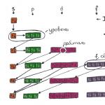
In 1%, the virus overcomes the blood barrier and spreads
through brain tissue, damaging large
motor cells in the anterior horns of the SC and nuclei
medulla oblongata and pons until their death.
This leads to the development of flaccid muscle paralysis
limbs, torso, neck, intercostal muscles –
paralytic form Periods of illness:
Incubation (2-35 days, usually 7-14)
Pre-paralytic (2-5 days)
Paralytic (4-7 days)
Restorative
Residual effects
Classification
TypeForms
gravity
Flow
Typical with NS involvement:
non-paralytic
(meningeal)
Lightweight
Medium-heavy
Heavy
Uncomplicated
Complicated
paralytic 0.1-1%
(spinal, bulbar,
pontine, mixed)
Atypical:
abortive (visceral)
subclinical
inapparent
(virus carrier) Clinic
Non-paralytic forms:
They are rarely diagnosed, only in epidemic outbreaks.
Abortive form (99% of all diseases) - acute onset, fever,
moderate intoxication, malaise, weakness, headache.
Catarrhal phenomena in the upper respiratory tract - slight cough, runny nose. Signs
gastroenteritis or enterocolitis (abdominal pain, dysfunction
intestines). Fever persists for 3-7 days, but after 2-3 days it is possible
repeated rise of T. Notice the pronounced sweating in
head and neck areas. Full recovery.
Meningeal form - serous meningitis against the background of catarrhal
phenomena from the VDP. Kernig's, Brudzinski's signs, rigidity
neck muscles, severe headache, insomnia, nausea, sometimes
vomit. Sometimes accompanied by pain in the limbs, back,
skin hyperesthesia, pain on palpation of nerve trunks,
horizontal nystagmus. Patients are adynamic, reluctant to sit down,
while leaning on his hands (tripod symptom). Moderate in cerebrospinal fluid
an increase in lymphocytes with normalization in the 3rd week of the disease.
It may be difficult, but the outcome is favorable.
Encephalitic
More often in young children. With focal neurological symptoms,
but without paralysis. Paralytic forms occur in unvaccinated children and occur
in 4 stages:
Preparalytic - acute increase in T, catarrhal symptoms with
sides of the upper respiratory tract (rhinitis, tracheitis, bronchitis) and intestinal dysfunction
(loose stools, vomiting, anorexia). Then T returns to normal, and h/w 2-4 days
rises again to 39-40 (two-wave character), headache appears
pain, drowsiness, sweating, spinal symptoms (pain with
any bending of the limbs, turns of the body, the child cannot
bend to reach his knees with his face, cannot sit without support, and
if he can be seated, he sits with support on his hands behind his back - sm
"tripod"; characteristic "potty" - the child resists and
cries in pain when being put on the potty).
The face is pale, amicable, the eyes are lifeless, the cheeks and lips are
cyanotic hue. Pain in the back, along the nerves, twitching in
individual muscle groups, tonic and clonic convulsions.
Sweating is pronounced. Hyperesthesia is pronounced: the child does not give
cover himself with a blanket, demands to take off his clothes. Adynamia (on request
the child refuses to perform any movement or does it with
labor. Lasts 3-5 days. Paralytic stage - symptoms of intoxication
are growing. Headache, repeated vomiting,
meningeal symptoms, T. After 5-7 days from
the onset of the disease develops suddenly
paralysis, flaccid, with low muscle tone,
hyporeflexia. Usually found in the morning
(morning paralysis). Affected limbs
cold, pale, cyanotic. To this moment
T is normalized, intoxication disappears.
The legs, pelvic girdle muscles, and
lesions of the intercostal muscles and
diaphragm. Muscle contractures develop and
joint deformities. The recovery period begins with
normalization of functions of easily affected
muscles, goes slowly. Deeply affected
muscles do not recover. By the end of the 1st
months, atrophies develop, which in
further progress. Expressed
vegetative disorders: cold snap
limbs, cyanosis, impaired
sweating Recovery is underway
during the 1st year, then slows down.
Developed paralysis, as a rule,
do not disappear completely.
Constant supervision by a neurologist is necessary,
orthopedist and exercise therapy doctor. Complications:
Pneumonia
Atelectasis
Myocarditis Treatment
Mandatory hospitalization and treatment until persistently positive
dynamics of lost functions, at least 3-4 weeks.
Strict bed rest, heat on the affected limb.
Orthopedic regimen depending on the location of paralysis. In case of defeat
of the lower extremities, bolsters and feet are placed under the knee joints
rest at an angle of 90 degrees against the foot end of the mattress. In case of defeat
the upper limbs are held in a slightly abducted position.
Physiological position of the limbs using plaster splints,
Bed with a hard mattress, shield, prevention of bedsores.
In the acute phase, detoxification and dehydration therapy
(diacarb, furosemide), pain relief (analgin)
If necessary, mechanical ventilation.
For swallowing problems, feeding through a tube.
During the recovery period, dibazole, glutamic acid, vitamins
group B, nerobol, ATP, nicotinic acid, cerebrolysin, exercise therapy,
massage, paraffin, hot wraps.
Sanatorium-resort treatment 6 months after the acute period.
Operative and orthopedic care. Prevention
Isolate the patient for at least 40 days from the onset of the disease.
Quarantine for 21 days, all contacts under 15 years old, adults from the outbreak and
emergency single immunization live for those working in kindergartens
vaccine. If there are contraindications for it and for children under 4 years of age, immunoglobulin intramuscularly 3.0 ml. Observation with thermometry.
After hospitalization - final disinfection with
chlorine-containing products. Dishes, care items, toys are washed
using detergents. Bed and underwear
are boiling. The pot is treated with a concentrated solution
bleach.
Routine immunization with a live vaccine of three strains of the virus.
Since cases of vaccine-associated polio are possible (1
case per 1-2.5 million vaccinated people) associated with the 1st vaccination, in many
In countries, vaccinations are carried out with an inactivated vaccine. In our
country, the first 3 vaccinations are given with an inactivated vaccine, then
continue vaccination with live vaccine.
A program to eliminate the infection in the world is being implemented. Yekaterinburg and
The Sverdlovsk region had the status of a territory free from
polio, before the importation of polio from Tajikistan.
A room with “iron lung” devices, which house patients with respiratory paralysis, in one of the hospitals in California, California 1953

POLIO
Poliomyelitis –
(from the Greek polios - gray and myelos - bone or spinal cord) - an acute infectious disease (old name - “infantile paralysis”) caused by one of the smallest viruses - poliovirus from the family of picornaviruses, which do not have an envelope and contain RNA

POLIO
The duration of the incubation period is from 4 to 30 days
Polio is transmitted through contaminated hands, food, water
It is assumed that the infection can also be transmitted by airborne droplets
The virus affects the central nervous system, causing a degenerative-inflammatory process in the anterior horns of the spinal cord and the gray matter of the subcortex

POLIO
ICD CLASSIFICATION – 10
A.80. Acute polio
A.80.0. Acute paralytic poliomyelitis associated with vaccine virus.
A.80.1. Acute paralytic poliomyelitis caused by wild imported poliovirus (types 1, 2 or 3).
A.80.2. Acute paralytic poliomyelitis caused by wild local (endemic) poliovirus (types 1, 2 or 3).
A.80.3. Acute paralytic poliomyelitis, other and unspecified etiology.
A.80.4. Acute non-paralytic poliomyelitis

POLIO
Clinical forms of paralytic poliomyelitis
2. Bulbar
3.Pontina
4.Mixed
(bulbospinal,
pontospinal)
Motor nuclei of the cranial nerves of the brainstem
Isolated lesion
nuclei of the facial nerve in the area of the Varoliev bridge
Damage to the nuclei of the cranial nerves and spinal cord

POLIO
The most common form.
In the pre-vaccination period it was 46-54%

POLIO
Spinal form of acute polio
Preparalytic period
The onset is acute, with high T° and intoxication
Catarrhal phenomena, loose stools
Children are lethargic, moody, have poor sleep and appetite
Headaches, pain in the back, neck, limbs
On examination, meningeal signs and tension symptoms
Pain when sitting up in bed with legs extended
Myoclonus of muscles or muscle groups in areas of future paresis

POLIO
Spinal form of acute polio
Paralytic period
The lower extremities are most often affected
Paralysis is flaccid in nature
Severely limited range of motion
Low tone
SR low or absent

POLIO
Spinal form of acute polio
Features of paralysis:
short rise time
the proximal parts are more often affected
asymmetrical, “mosaic” arrangement
sensory, pelvic disorders and pyramidal symptoms are absent

GLOBAL POLIO ERADICATION INITIATIVE
In 1988, the Forty-First World Health Assembly, then composed of delegates from 166 Member States, adopted a resolution to eradicate polio from the world
This marked the beginning of the Global Polio Eradication Initiative, led by the World Health Organization, Rotary International, the US Center for Disease Control (CDC) and the United Nations Children's Fund UNICEF
The Initiative was adopted following the confirmation of smallpox eradication in 1980 and built on the progress made in eradicating poliovirus in the Americas in the 1980s
Poliovirus Discovered by Enders, Weller and Robbins Discovered by Enders, Weller and Robbins Poliovirus, genus of enteroviruses Poliovirus, genus of enteroviruses RNA-containing RNA-containing 3 serotypes 3 serotypes Source of infection and reservoir of the virus - humans Source of infection and reservoir of the virus - humans Isolated from the nasopharynx ( 1-2 weeks) and intestines (several weeks) Isolated from the nasopharynx (1-2 weeks) and intestines (several weeks)

Epidemiology Airborne and alimentary routes of infection Airborne and alimentary routes of infection Resistance of the virus Resistance of the virus Summer, early autumn Summer, early autumn More often in southern countries More often in southern countries Children under 5 years old Children under 5 years old


Clinical forms Inapparent Inapparent Abortive (visceral) Abortive (visceral) Meningeal Meningeal Paralytic (“infantile spinal palsy”) Paralytic (“infantile spinal palsy”) spinal, pontine, bulbar, mixed spinal, pontine, bulbar, mixed

Course of paralytic forms Preparalytic period (1-2 days) Preparalytic period (1-2 days) Paralytic period (in the first hours after a decrease in temperature, morning paralysis) - up to 2 weeks Paralytic period (in the first hours after a decrease in temperature, morning paralysis ) - up to 2 weeks Recovery period Recovery period Period of residual effects Period of residual effects Mortality - 10%, disability - 40% (at the beginning of the 20th century).

Diagnostics Treatment Cellular-protein dissociation in the cerebrospinal fluid Cellular-protein dissociation in the cerebrospinal fluid Virological study (nasopharyngeal swabs, feces) Virological study (nasopharyngeal swabs, feces) Serological study in dynamics Serological study in dynamics Rest Rest Dehydration Dehydration Intensive therapy Intensive therapy Prevention of contractures Prevention of contractures Rehabilitation treatment Rehabilitation treatment

Isolation 3 weeks Isolation 3 weeks Observation of contacts Observation of contacts 3 weeks contacts 3 weeks Since 1959 - trivalent OPV vaccine at 1, 2 and 7 years Since 1959 - trivalent OPV vaccine at 1, 2 and 7 years creating herd immunity - 90% of the world's population needs to be vaccinated. creating herd immunity - it is necessary to vaccinate 90% of the world's population. Prevention

Every year, paralytic poliomyelitis in the countries of the WHO European Region fell ill. - 28.5 thousand children. – 7.7 thousand children – 7.7 thousand children 1975 – 1.1 thousand children 1975 – 1.1 thousand children 1979 – 0.2 thousand children 1979 – 0.2 thousand children

Sabin vaccine (OPV) - 1959 - oral pomyomyelitis vaccine - live attenuated polio virus (so-called vaccine strains). Administered through the mouth, drop by drop. Induces persistent powerful immunity, including in the intestines. Vaccine-associated polio is very rare - 1 in 5 million vaccinated people.

Salk vaccine (IPV), Denmark - inactivated, administered by injection, does not cause complications, induces good general immunity, but does not develop local protection in the intestine.