Uterine fibroids - what is it and why is it dangerous? Symptoms and treatment
This is a benign, hormonally controlled hyperplasia of the muscular layer of the uterine wall of mesenchymal origin, which develops as a result of somatic cell mutation that occurs under the influence of various damaging factors.
It is presented in the form of a tumor consisting of intertwined muscle and connective tissue fibers. In clinical practice, fibroids of the uterine body and cervical fibroids occur.
What it is?
Uterine fibroids are a benign tumor that arises in the muscular layer of the uterus - the myometrium. It is one of the most common diseases in women, reaching an incidence of 12 - 25% of all gynecological diseases.
The highest incidence of uterine fibroids occurs in the late reproductive period and before menopause. There is an opinion that the true prevalence of fibroids is much higher and reaches more than 70% both in Russia and abroad.
Causes of uterine fibroids
Myoma is a polyetiological disease, the following risk factors may play a role in its development:
- hormonal imbalance;
- inflammatory pathology of the female genital area;
- the use of intrauterine contraceptives, for example, spirals;
- complicated course of labor;
- performing abortions;
- adenomyosis;
- obesity;
- disease of the thyroid gland, adrenal glands, organs of the hypothalamic-pituitary system.
Myoma is a hormonal-dependent tumor, evidenced by the following facts:
- Receptors for sex hormones are found in the tumor tissue;
- as a rule, after the onset of menopause and a sharp decline in the level of sex hormones, tumors undergo reverse development;
- Most often, fibroids appear in women of reproductive age, whose bodies have a high content of estrogen;
- After menopause, fibroids may appear in women who take estrogen-containing medications.
Classification
Depending on the location of fibroids, there are several terms in clinical practice:
- Pedunculated uterine fibroids are not a separate gradation, since pedunculated fibroids can have submucosal and subserous fibroids. Within the definition, the size of the pedicle, fibroids on a wide or narrow base can be noted.
- Interstitial or intramuscular fibroids - a neoplasm located in the muscular wall of the uterus.
- Submucosal or submucosal fibroid - a tumor grows right inside the uterus under the mucous membrane and extends into the lumen of the uterus.
- Subserosal fibroids - the neoplasm is located on the outer surface of the uterus and is separated by a membrane that separates the uterus and abdominal organs.
The first signs of uterine fibroids
In the initial stage, small uterine fibroids are not accompanied by noticeable symptoms. It can be detected during the next gynecological examination.
As uterine fibroids grow, the first signs may appear:
- long, heavy and irregular menstruation;
- constipation;
- infertility;
- bleeding;
- anemia;
- frequent urination;
- heaviness and constant pain in the lower abdomen;
- bleeding during sexual intercourse;
- lower back pain;
- abdominal enlargement not associated with significant weight gain;
- frequent miscarriages.
Why is pedunculated uterine fibroid dangerous? When the “leg” is twisted, inflammation and rupture of the tumor occurs. This causes severe bleeding, severe pain in the lower abdomen and fever. This condition can be fatal.

Symptoms
When the nodes are large, the function of neighboring organs is impaired: constipation, disturbances in bladder emptying, and frequent urination. In some cases, tumor nodes can cause compression of the ureter with further disruption of the outflow of urine from the kidney, which leads to the death of the kidney. Symptoms of compression of neighboring organs more often appear with large sizes of nodes and low localization of nodes.
- Interstitial uterine fibroids (with intermuscular nodes) lead to longer, heavier and more painful menstruation. Especially often, heavy, prolonged and painful menstruation is observed with a combination of uterine fibroids and adenomyosis. Also, with intermuscular growth of the node, part of it can grow towards the uterine cavity. With this localization of fibroids, the same symptoms are observed as with submucous growth of the node, and their severity depends on the size of the submucosal fragment of the node.
- For the submucosal location of the node (submucous fibroids), heavy menstruation is most typical, and with large sizes, when the node begins to occupy a large area of the uterine cavity, bleeding from the genital tract loses its cyclicity, and sometimes does not stop at all. With this location of the node, the patient almost always develops anemia due to heavy bloody discharge from the genital tract. Submucous fibroids can cause cramping pain, most often occurring during menstruation. The submucosal node is a kind of “foreign” body for the uterus, from which it is trying to free itself. Sometimes such nodes can even be “born” on their own. This process is accompanied by very severe cramping pain and bleeding.
- Subserous (subperitoneal) fibroids often manifest clinically as painful symptoms. The pain is localized in the lower abdomen and/or lower back. Their appearance is associated with tension in the ligamentous apparatus of the uterus and the pressure of growing fibroids on the nerve plexuses of the pelvis. If blood circulation in the node is impaired, the pain is acute and very intense.
Pain accompanies the development of fibroids in every third woman and can have different origins. With small interstitial nodes, painful menstruation is observed. Constant aching pain is observed with pronounced growth of nodes. With necrosis of the node, the pain syndrome is pronounced, an increase in body temperature and intoxication may occur. Also, an emergency situation may arise if the patient has pedunculated subserous nodes. When the “leg” is thin, the node is torsed; The power supply in the node is disrupted. This situation often manifests itself as an acute condition: severe pain, with the possible development of peritonitis. In such a situation, urgent surgical intervention is required.
Diagnostics
The diagnostic process for suspected myomatous formation involves the following measures:
- Anamnesis collection. Particular importance is given to the age criteria of the patient, since such myomatous pathology is found mainly in women of reproductive age. As a rule, women have complaints regarding menstruation, which can become longer, heavier, painful, and irregular.
- Ultrasound diagnostics of the pelvic organs helps to accurately identify myomatous formation, determine its parameters and location.
- Gynecological examination. It is carried out on a gynecological chair using the necessary instruments. The size of the uterine body, the location of the ovaries, the shape and mobility of the cervix, etc. are taken into account.
- Diagnostic curettage. Usually performed to determine endometrial changes, as well as to exclude cervical or uterine cancer.
- Hysterographic study. Hysterography refers to radiopaque techniques that allow you to visually see the uterine cavity. To do this, the patient is injected with a contrast agent into the body of the uterus, and then an image is taken.
- Laparoscopy. A similar technique refers to surgical methods. The laparoscope is inserted through punctures in the peritoneum and displays an image of the organ being studied on the monitor. During the procedure, it is possible to obtain biomaterial for histology or removal of small tumors, etc.
- Hysteroscopic examination. Helps visualize the uterine cavity. It is carried out using special equipment that is inserted into the uterus. This method is not only diagnostic, but also therapeutic. In this way, various polyps are removed and biomaterial is obtained for histological examination.
The approach to the treatment of uterine fibroids is determined in accordance with the stage of formation.
How to treat uterine fibroids?
There are two main methods of treating uterine fibroids:
- Conservative treatment – with the help of medications and non-invasive procedures.
- Surgical treatment - through surgery.
The choice of treatment method depends on the severity of the clinical symptoms of uterine fibroids, the size of the fibroid node, the woman’s age, and the intention to have children in the future.
The conservative method is based on the use of hormonal drugs orally and by injection, as well as symptomatic therapy (pain relief, treatment of anemia, reduction of blood loss, etc.). Conservative therapy is effective only in controlling tumor growth. It is impossible to eliminate uterine fibroids conservatively. Therefore, the non-surgical treatment method is used mainly in women of advanced childbearing age.
At the same time, tumor growth is inhibited until the onset of menopause. At this time, there is a high probability of spontaneous resorption of the tumor.

Treatment of fibroids without surgery
Non-surgical treatment of uterine fibroids is a set of measures, including adherence to an appropriate diet, the use of immunomodulators, herbal medicine, approved physiotherapeutic procedures, and the use of hormonal medications.
The course of treatment consists of the following stages:
- Anti-inflammatory therapy for infectious processes in the gynecological area;
- Activation of the immune system with special drugs;
- Adjustment of diet and diet;
- Normalization of the endocrine system;
- Formation of an even psycho-emotional background;
- Elimination of bleeding;
- Treatment of anemia;
- Bringing the menstrual cycle back to normal.
At home, treatment of uterine fibroids without surgery involves the use of the following medications:
- Antiprogestogens. Mifepristone (RU-486) is often used as preparatory therapy before surgery or for the treatment of fibroids. This drug not only eliminates the symptoms of the pathology, but also helps reduce the size of the formation.
- Gonadotropin-releasing hormone agonists. Drugs in this group include: triptorelin - (Decapeptyl, Diferelin, Decapeptil Depot), buserelin, goserelin (Zoladex), leuprorelin (Lucrin Depot). The disadvantage of using these drugs for the treatment of uterine fibroids is that after the end of therapy, the nodes can be restored in size, and long-term use of these drugs is not permissible, as it can lead to the development of osteoporosis and other consequences for the female body due to a lack of estrogen. Within six months of using these products, the size of fibroids can be reduced by 50%, and the intensity of fibroid symptoms can also decrease. These medications are often used to shrink uterine fibroids before surgery to reduce blood loss during surgery.
- Combined oral contraceptives. These include the following combinations: desogestrel with ethinyl estradiol (Mersilon, Marvelon, Novinet), ethinyl estradiol with norgestrel (Ovidon, Rigevidon). These remedies effectively help reduce the symptoms of uterine fibroids, such as pain in the lower abdomen and bleeding. However, for the treatment of uterine fibroids in order to reduce its size, tablets of this group are not a priority, since they do not always lead to a reduction in nodes. Under the influence of such therapy, only nodes whose initial size did not exceed 1.5 cm can shrink in size.
- Gestagens. The use of such drugs today is a controversial issue, since some doctors believe that the use of such drugs, and especially dydrogesterone (Duphaston) for uterine fibroids, is unacceptable. Others believe that the use of this drug is completely justified, since it is the lack of progesterone that causes the growth of fibroids. The effectiveness of therapy with drugs such as linestrenol (Orgametril, Escluton), medoxyprogesterone acetate (Provera, Depo-Provera), nor-ethisterone (Norkolut, Primolut-Nor) for uterine fibroids has not been proven. , so many gynecologists do not recommend these drugs. However, the use of these agents in cases of combination of endometrial hyperplasia and uterine fibroids is justified. The growth of fibroids occurs not simply from a deficiency or excess of any hormone, but rather from an imbalance between hormones, so the use of such drugs should not be carried out everywhere, but only when indicated.
- Antigonadotropins. Danazol (Vero-Danazol, Danazol, Danoval, Danol, Danogen), gestrinone (Nemestran). For uterine fibroids, treatment with these drugs is rarely used, since they cannot reduce the size of the nodes, but only help reduce the intensity of the symptoms of fibroids; moreover, they have a number of undesirable side effects, such as acne, voice changes, and hair growth on the body and face. These medications for fibroids can only be used if other treatments are ineffective.
FUS ablation of fibroids
A method of treating fibroids based on heating the node tissue with high-intensity focused ultrasound, due to the transfer of a large amount of energy to a clearly localized area, after application of which destruction of the node tissue occurs - thermal necrosis. The principle of operation is very similar to the concentration of sunlight with a magnifying glass. The pinnacle of development of the method today is the combination of a focused ultrasound source (emitter mounted in the table) and an MRI machine. Ultrasound is performed through the anterior abdominal wall. The MRI scanner controls the localization and, most importantly, the heating temperature in real time.
The method allows destruction of a clearly limited area without damaging healthy tissue. The zone between the “treated” and healthy tissue is only a few rows of cells. Thus, focused ultrasound today has come closer than ever to the concept of “ideal surgical instrument.” The procedure does not require the introduction of instruments into the body cavity, anesthesia and hospitalization, but it is not possible in all cases and requires proper patient selection. The substrate for the effects of focused ultrasound is the connective tissue located in the fibroids.
Connective tissue accumulates energy well and allows it to reach the temperature necessary for thermal necrosis. Thus, so-called fibroids, which make up 70% of all fibroids, respond very well to treatment using FUS. Leiomyomas, or cellular fibroids, are absolutely not suitable for treatment with the FUS method due to the lack of a substrate for exposure and a high-intensity blood supply, which does not allow sufficient heating of the tissue.
During the period of testing the method and its introduction into clinical practice, improper selection of patients caused frequent relapses and was mistakenly interpreted as the ineffectiveness of the method. To assess the possibility of performing FUS ablation, the patient must undergo an MRI, which determines the type of fibroid. Recently, numerous works by various authors, based on retrospective studies of treated patients, have shown the high effectiveness of the method, which is not inferior in terms of performance to other methods of treating uterine fibroids, provided that it is used correctly.
Indications for the use of FUS ablation for uterine fibroids are standard indications for organ-saving treatment for uterine fibroids. Depending on the clinical tasks set by the gynecologist, the technology can be used in four tactical options. 1. Organ-saving treatment of uterine fibroids. 2. Prevention of clinical manifestations of uterine fibroids (which allows you to abandon the expectant outpatient practice regarding the growth of fibroids). 3. Preparation of the fibroid node for transcervical myomectomy (as a result of FUS destruction, the size of the node and the intensity of blood flow are reduced, which significantly reduces the risk of possible complications and facilitates the operation); 4. In order to delay surgical treatment.
Unlike other methods, FUS-MRI ablation of uterine fibroids is a non-invasive, organ-saving method, without hospitalization, without pain, it gives a woman the highest chance of a successful pregnancy compared to other methods, and has a minimal percentage of complications among all methods used for treating uterine fibroids (currently less than 0.05%). The method has received recognition in many countries CE (Europe), FDA (USA), MHLW (Japan), CFDA (China), ANVISA (Brazil), Canada FDA, KFDA (Korea), Russian Federation, Taiwan.

Uterine artery embolization (UAE)
This is the introduction of special plastic balls into the uterine arteries in order to stop the blood supply to certain areas of the uterus and fibroids. The UAE procedure has very limited indications for use and is used mainly in women over 40 years of age with submucosal myomatous nodes and severe uterine bleeding.
The use of uterine artery embolization methods in women of reproductive age, according to the recommendations of the European and American Societies of Obstetricians and Gynecologists, is prohibited. There are a huge number of publications in the literature illustrating the consequences of unsuccessful uterine artery embolization. As a rule, these are young nulliparous women aged 25–35 years who lost the opportunity to have children as a result of the UAE procedure for small fibroids that did not require treatment at all.
When is surgery to remove the uterus for fibroids indicated?
Indications for radical surgical treatment of uterine fibroids:
- Tumor size from 12 weeks (a voluminous tumor compresses neighboring organs and blood vessels, interfering with their normal functioning);
- Rapid growth of myomatous node (from 4 weeks per year);
- Myoma causes massive bleeding;
- Severe pain syndrome;
- Torsion of the leg and necrotization of the myomatous node;
- Birth of a submucosal myomatous node;
- Combination of endometriosis and fibroids;
- Suspicion of malignancy of fibroids.
Depending on the size, location and type of fibroids, the doctor selects the most convenient type of operation to eliminate it. Myomectomy today is performed in three ways:
- laparoscopy - through a small hole in the abdomen;
- through the vagina using a special instrument - hysteroscopy;
- strip surgery - open, through an incision in the lower abdomen (very rare);
- if gentle operations are not possible, and it is impossible to stop the growth of fibroids by other means, treatment involves complete resection of the uterus - hysterectomy.
Hysterectomy and laparotomy are the most popular surgical methods, as they have a number of advantages: rapid recovery after surgery, preservation of the ability to become pregnant and bear a child in the future, and almost complete absence of traces from the operation.

Prevention
Prevention of uterine fibroids consists of following a diet, sleep and rest patterns, and reducing stressful situations. An important method of preventing uterine fibroids is timely pregnancy and childbirth (up to 25 years), as well as preventive examinations by a gynecologist.
Uterine fibroids- a chronic disease of the uterine body, manifested by the growth of one or several tumor-like nodes in its muscular layers. As a result, the size of the uterus increases and its shape becomes deformed. This leads to unpleasant symptoms - pathological bleeding, frequent urination, pain in the lower abdomen and pelvic region, and reproductive disorders. But this doesn't always happen - signs of uterine fibroids in women depend on the number of nodes, as well as their size. In more than half of the cases, myomatous nodes are small in size and do not manifest themselves in any way.
The disease is very common. According to statistics, by the age of 45, up to 70-80% of women suffer from fibroids.
Treatment tactics depend on the stage of the tumor, the speed of its growth, and the age of the patient. Includes conservative and surgical methods. An asymptomatic course requires only regular monitoring by a gynecologist.
Causes of the disease
Myoma is a hormone-dependent disease. This means that the nodes grow in response to the influence of female sex hormones, in particular progesterone and estrogen. Therefore, it occurs only in women of reproductive age. However, it most often develops in the late reproductive and premenopausal periods. With the onset of menopause, small and medium-sized nodules tend to regress, or, as people say, dissolve. In women after menopause, fibroids may appear in response to taking estrogen-containing drugs.
There is no scientific data that would reliably confirm that the cause of the development of fibroids is a hormonal disorder. In many patients, nodes appear against the background of normal hormonal parameters. This means that this hormone-dependent disease is not hormone-related.
Some hereditary dependence was identified, but it is also impossible to talk about the direct influence of the hereditary factor of this disease.
There are risk factors:
- later menarche;
- a large number of abortions;
- heavy menstrual flow;
- inflammatory diseases of the genital area;
- ovarian cysts;
- long-term ill-chosen contraception;
- liver dysfunction;
- absence of childbirth and lactation by the age of 30;
- excess weight;
- carbohydrate metabolism disorders;
- sedentary lifestyle;
- stress.
However, today it is impossible to reliably determine the cause of fibroids and the likelihood of its development in a particular woman.
Classification of myomatous formations
The body of the uterus consists of a muscular layer and a mucous membrane that lines its inside.
Myomatous formations are localized in muscle connective tissue. The source of their growth growth is one defective cell, which undergoes certain changes and begins to divide at a faster rate than neighboring ones.
As a result, a myomatous node appears - a local accumulation of chaotically intertwined smooth muscle fibers. On average, its dimensions range from a few millimeters to several centimeters. However, sometimes very large tumors occur. Some reach a weight of several kilograms.
Myomatous cells practically do not degenerate into malignant ones. This happens in less than 1% of cases.
Attention! Even the rapid growth of fibroids is not a sign of its malignancy

Fibroids of the uterine body can be located in different layers. Based on these characteristics, nodes are divided into the following types:
- Submucous or submucosal - grows into the uterine cavity. It can protrude inward completely, half or less.
- Intramural or intermuscular - located inside the wall of the organ.
- Subserous or subperitoneal - protrudes outside (into the peritoneum).
Among these types there is fibroid, which grows on a stalk.
Symptoms of uterine fibroids
Although the disease is highly widespread, about 35-40% of patients experience severe symptoms; the rest are asymptomatic.
Manifestations of fibroids:
- Heavy and prolonged menstruation.
- Clots in menstrual blood.
- Feeling of pressure in the bladder area.
- Frequent urination.
- Pain in the lower abdomen.
- Abdominal enlargement.
- Pain during sexual activity.
With large tumors, complications arise.
Common ones include the development of anemia caused by heavy and prolonged menstrual bleeding.
Reproductive function disorder - usually occurs when the node is located intramurally. In this case, difficulties arise with the onset of pregnancy, since the nodes prevent the fertilized egg from attaching to the uterine mucosa. It can also cause miscarriage.
A rare complication is dysfunction of nearby organs: the bladder and rectum.
Diagnostics
A doctor may suspect uterine fibroids based on the patient’s complaints, and after a gynecological examination, if the size of the organ increases.
Previously, the degree of development of the disease was correlated with weeks of pregnancy, based on how much the patient’s uterus was enlarged. This has been the case since the days when there was no ultrasound diagnostics. Now the definition of the type “fibroids corresponding to 8-9 weeks of pregnancy” serves as a marker of the size of fibroids, but not a diagnostic criterion.
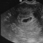
The disease can only be reliably diagnosed using the results of an ultrasound examination. Ultrasound imaging allows you to accurately determine the size of the nodes, their location and type. When reporting the results, it is necessary that the ultrasound specialist make not only a description of the nodes, but also a schematic drawing of the uterus with their location and an indication of the size of each.
This is important for the attending physician when planning further treatment. For example, to understand exactly how to remove tumors. Or is it possible for a patient to become pregnant with this arrangement of nodes.
Since ultrasound cannot always cope with formations that are too large and a very enlarged uterus, in some cases an MRI is prescribed in addition to ultrasound.
Hysteroscopy is one of the additional diagnostic tools. This is an endoscopic examination, when a probe with a camera is inserted into the uterine cavity and it is examined from the inside. The need for this method does not arise often.
Treatment of uterine fibroids

Uterine artery embolization (UAE)
This is a minimally invasive and fairly safe method, which consists of blocking the blood flow to the myomatous nodes. As a result, they receive no nutrition and regress within a few hours.
In this case, no cuts are made. A substance containing emboli - small balls that, like a plug, block the blood flow - is injected into the femoral artery using a tube.
This is a completely organ-preserving method. The rate of serious complications with its use is about 1%.
There are pronounced adverse reactions. These are painful sensations in the lower abdomen for several hours that require relief with an anesthetic drug. Nausea, vomiting, fever, and difficulty urinating may occur for several days.
The method is indicated for women of reproductive age with fibroids that have severe symptoms.
Surgical methods
The largest number of surgical operations in the field of gynecology are performed on uterine fibroids. The nodes on the uterus and the uterus itself are surgically removed if other types of treatment have been ineffective.
Myomectomy
Removal of myomatous nodes with preservation of the organ.
The method of operation usually depends on the type of nodes:
- Hysteroscopic. In this case, no incision is made. Removal is performed with a resectoscope, which is inserted through the vagina. This type of operation is possible only when the nodes are located submucosally.
- Laparoscopic. The operation is performed using a laparoscope through several punctures in the abdominal cavity.
- A bdominal. Incisions are made in the abdomen and uterus. This type of operation is used for multiple fibroids. Although this is the most invasive intervention with a long recovery period, it also belongs to organ-saving operations.
The most appropriate is to remove macroscopic myomatous formations of medium size (from 2 to 5 cm in diameter) before they grow to large sizes. This makes their treatment much more difficult and may lead to the need for a hysterectomy.
Hysterectomy (complete removal of the uterus)
An extreme measure that should be resorted to only in case of severe symptoms and very large organ sizes, as well as if other methods do not produce results.
The operation is also performed using laparoscopic, hysteroscopic or abdominal methods.
Typically used for women over 40 who already have children.
All of these types of surgical interventions are performed under general or epidural anesthesia.
Drug therapy

Contraceptives. They can reduce the amount of bleeding and temporarily stabilize the growth of small nodes.
Drugs that cause temporary menopause. During use (usually 6 months), the nodes decrease, but after discontinuation they quickly grow to their previous, and sometimes to larger sizes. Complete regression of fibroids is only possible if they are very small.
Mirena intrauterine device. Thanks to its effect on the endometrium, it removes the symptom - heavy menstrual bleeding.
The listed drugs are symptomatic and do not have a lasting therapeutic effect.
Other methods
Cryotherapy(freezing of nodes).
FUS ablation(remote evaporation with a narrowly directed ultrasonic beam).
These are new methods with little evidence. Therefore they are not widely used.
Attention! Dietary supplements, including indole, leeches, reflexology, herbal medicine, do not treat uterine fibroids.
Modern approaches to treatment
Which methods the doctor will use depends on many factors.
Modern concept of treatment of uterine fibroids:
- Uterine fibroids require treatment only if indicated.
- Therapy should not be more severe than the disease itself.
- The approach to treatment should be as organ-preserving as possible.
- Treatment should provide the most lasting effect.
- Focus on preserving reproductive function even if a woman is not planning a pregnancy now.
Symptoms and treatment of fibroids interconnected. When choosing therapy, you need to answer the following questions:
- Does fibroid interfere with a woman’s life?
- What nodes were found and where exactly.
- Are they growing?
- Is the woman planning pregnancy and when?
- Patient's age.
Since fibroids do not degenerate into cancer, and in approximately 60% of cases they do not manifest themselves in any way, or the symptoms are insignificant, a woman should not be subjected to treatment, much less surgery. The recommendations of some doctors to remove fibroids just because they exist are completely unjustified, not to mention removing the entire organ. The patient’s health may suffer more from radical treatment than from small fibroids.
However, if the nodes grow steadily from measurement to measurement, this is also a reason for treatment.
But if a woman is premenopausal, and the growth of fibroids and symptoms are moderate, the doctor may choose a wait-and-see approach.
After the onset of menopause, the production of sex hormones stops and the fibroids degrade. If your periods are heavy, the Mirena coil may be prescribed as a symptomatic treatment.
If a woman is planning a pregnancy, it is important to understand how much the nodes can interfere with this - will they deform the shape of the uterus, will they interfere with the attachment of a fertilized egg to its walls, will the rapidly growing nodes interfere with the development of the fetus. If these factors are excluded, it is possible to become pregnant with fibroids.
If neoplasms lead to infertility and miscarriage, treatment is necessary - embolization or myomectomy. It is important to take into account that fibroids are a disease with a likelihood of recurrence. Therefore, it is advisable to carry out such an intervention if a woman is planning a pregnancy in the near future. If she plans to put it off for several years, there is a chance that the nodes will grow back.
Prevention
To prevent the disease, it is important to exclude everything that puts a woman at risk - stress, anemia, venous congestion in the pelvic area, illiterate contraception, inflammatory processes, abortions.
Therefore, preventive measures will be as follows:
- normal sleep;
- proper nutrition;
- maintaining optimal weight;
- physical activity;
- regular sex life;
- timely treatment of diseases of the female genital area;
- maintaining pregnancy and breastfeeding for at least 4-6 months.
Also, to prevent fibroids, it is important to realize reproductive function in a timely manner. It is optimal if the first birth occurs at about 22 years old, and the second at 25 years old. Late first birth leads to premature aging of myocytes (muscle cells), which means a decrease in the ability of the uterus to stretch and contract, and a tendency to form nodes.
Uterine fibroids are not easy to correct, but they are not dangerous diseases. For fibroid therapy to be most effective, it is important to identify it in time. Therefore, preventive measures include regular visits to the gynecologist.
- Pain in a woman's lower abdomen
- Heavy menstruation
Uterine fibroids are a benign tumor in women of reproductive age (this disease mainly affects women between 30 and 45 years old). The tumor appears as randomly enlarged muscle fibers of the uterus, forming a knot that is densely entwined with altered vessels (their diameter is several times larger than normal vessels) that feed it. The peculiarity of the tumor is that its growth and development depends on the level of sex hormones in the woman’s body (hormone-dependent tumor).
The disease accounts for about 30% of all gynecological diseases and is found in 80% of women who may not experience any changes in their own health.
Uterine fibroids can appear in nulliparous young women, in older women, after gynecological operations, after childbirth, during menopause, and even during pregnancy.
In the case of hemorrhagic syndrome with uterine fibroids, as a rule, emergency surgical intervention is necessary, despite the fact that the patient’s condition is sometimes extremely severe and often aggravates existing homeostasis disorders due to possible blood loss during the intraoperative period.
Dimensions of uterine fibroids
Particular attention should be paid to the fact that the size of uterine fibroids has almost no determining influence on the symptoms of the disease itself (previously, uterine fibroids were determined by the enlargement of the uterus, as during pregnancy, hence the “fibroids at 18 or 20 weeks”). However, with the development of diagnostics, primarily the availability of ultrasound and MRI, this became a thing of history.
Currently, it is not the size of the tumor that plays a big role, but its type and location (for example, on the back wall). Large fibroids may not be felt by a woman at all (a woman may also not experience pain in the lower abdomen) and may not affect her well-being. At the same time, small fibroids in the submucosal layer of the uterus cause pain in the lumbar region, can disrupt the menstrual cycle, and provoke heavy menstruation even when carrying a child during pregnancy.
Classification of the disease
There are several classifications of this disease depending on the location of uterine fibroids, its cellular composition, and topographic location:
- Subserous uterine fibroids(the myomatous node is located in the uterus directly under the peritoneum covering the uterus). The fibroid node grows towards the abdominal cavity.
- Submucosal fibroid(the node is located, on the contrary, directly under the mucous membrane of the uterus). The node grows into the uterine cavity.
- Intermuscular uterine fibroids(the node develops in the thickness of the uterus).
- Intraligamentary(interligamentous), when nodular uterine fibroids develop between the broad ligaments of the uterus.
Currently, doctors more often use the clinical classification, which combines several others and is of greatest value in diagnosis and selection of further treatment tactics.
Within this classification there are:
- clinically insignificant fibroids or small uterine fibroids;
- small multiple fibroids;
- medium-sized uterine fibroids;
- multiple uterine fibroids with a leading medium-sized node;
- submucous uterine fibroids;
- large uterine fibroids;
- pedunculated uterine fibroids;
- complex uterine fibroids.
It is worth noting that 90% of all myomatous nodes are uterine fibroids. And the tumor itself has a tendency to develop multiple foci (according to scientists, the presence of only one node of uterine fibroids indicates only the initial stage of development of the disease).
Causes of uterine fibroids
Currently, the causes of uterine fibroids are not fully understood. All scientists agree that there is a precursor cell to education. However, scientists differ on the issue of the mechanism of its appearance.
One theory of the development of uterine fibroids involves the appearance of a genetic defect in smooth muscle cells during embryonic and subsequent development of the uterus due to a long and unstable period of embryonic changes. The second theory is the possibility of damage to the cells of an already mature uterus under the influence of various factors, which is confirmed by numerous studies (in a microscopic examination of preparations of the muscle tissue of the uterus (myometrium), myomatous nodes were found in 80% of cases).
According to modern concepts, uterine fibroids develop as follows. During multiple cycles of hyperplasia (an increase in the mass and volume of an organ without an increase in cellular elements) of the myometrium during the menstrual cycle, smooth muscle cells accumulate with a disturbed process of apoptosis (programmed death), which are exposed to various damaging factors: ischemia (insufficient blood circulation) caused by spasm arteries during menstruation, inflammatory processes, traumatic effects or endometriosis.
With each menstrual cycle, the number of damaged cells accumulates. Some cells are removed from the myometrium, while myomatous nodes with different growth potential are formed from others. The active growth bud in the first stages develops due to physiological fluctuations in hormone concentrations during the menstrual cycle. Subsequently, the resulting complex of cells activates local stimulating (growth factors) and supporting (local synthesis of estrogens from androgens) mechanisms, and the importance of the concentrations of sex hormones in a woman’s body for the formation of a myomatous node ceases to be decisive.
In addition, uterine fibroids are caused by disruption of the functioning of certain genes (HMGIC and HMGIY) involved in the rapid growth of embryonic tissues and located on chromosomes 12 and 6, respectively. A complete disruption of protein synthesis from these genes causes rapid cell division with the development of a malignant formation, while at the same time, its partial disruption is characteristic of various benign formations.
Thus, the complex of cells of the myomatous node, due to dysregulation of genes and the development of local mechanisms for activating and maintaining growth, constantly increases in size, while the cells of the unchanged myometrium are in a state of relative rest. Subsequently, the myomatous node increases the amount of connective tissue in its composition and intensifies the synthesis of estrogens from androgens, which leads to a slight decrease in the size of the formation, provided that it is deprived of hormonal stimuli.
Complications of uterine fibroids
The most common complication of uterine fibroids is malnutrition of the subserous node with subsequent necrosis of the node, less often - torsion of the pedicle of the subperitoneal node, acute bleeding, leading to persistent anemia of the patient. Such a complication as uterine inversion when a submucous node is born is extremely rare. Malignant degeneration is noted, according to some authors, up to 2%.
In 50% of cases, uterine fibroids can be asymptomatic. But, if you suspect uterine fibroids (see symptoms of uterine fibroids), you should immediately consult a doctor.
Diagnosis of uterine fibroids begins with detailed collection of information from the patient about his state of health (both past and present) and the factors that could influence it. Particular attention is paid to the number of births and their complications, the presence of abortions and their complications in the past. Separately, they find out about the presence of large and small intrauterine interventions (therapeutic and diagnostic curettage, placement and removal of intrauterine devices), the volume of menstrual bleeding.
Methods for diagnosing uterine fibroids
The first examination method is a bimanual (two-handed) vaginal examination. During this procedure, the doctor determines by touch the size and position of the uterus, and may also suspect the presence of uterine fibroids (if it is enlarged, the surface is uneven and tuberous, or the density of the uterus is heterogeneous).
Ultrasound examination of the pelvic organs for uterine fibroids is the second stage if this disease is suspected. This is a routine and widespread method for both the detection of uterine fibroids and its dynamic monitoring. Using ultrasound, the location of the nodes, their number and structure are determined. Such an ultrasound examination can be carried out in two ways: abdominal (the sensor is located on the anterior abdominal wall) and transvaginal (the sensor is inserted into the vagina).
In some cases, when a more precise determination of the location of uterine fibroids or clarification of its structure is necessary, magnetic resonance imaging may be required.
Hysteroscopy (examination of the uterine cavity using special instruments) and hysterosalpingography (determining the patency of the uterine cavity and fallopian tubes using special solutions using ultrasound or fluoroscopic examination) have retained their diagnostic value.
Important diagnostic methods are also the determination of a woman’s hormonal levels (levels of FSH, LH, prolactin, estrogens, progesterones, adrenal and thyroid hormones). A biopsy of the endometrium (the inner layer of the uterine cavity) to exclude its diseases, a smear to identify pathogens that are sexually transmitted, the state of the blood coagulation system and hemoglobin level are the necessary diagnostic minimum if uterine fibroids are suspected.
If there are great difficulties in diagnosing the disease, laparoscopy may be indicated for some women.
If uterine fibroids are suspected, the doctor consistently excludes the following diseases that can cause similar symptoms: benign and malignant voluminous diseases of the ovaries, endometrium, malignant diseases of the uterus (leiomyosarcoma), metastases of other tumors - and pregnancy itself.

The abdominal cavity can be opened using a midline incision or a transverse suprapubic incision according to Pfannenstiel. The surgical wound should be well diluted with a retractor, and the intestines should be delimited with napkins.
Removing a myomatous node on a “pedicle” does not present any difficulties and boils down to excision of the “pedicle” at its base. In this case, you should not make deep incisions in the direction of the body of the uterus, delving into the myometrium. It is enough to make an incision in the peritoneum and a thin layer of muscle fibers passing from the body of the uterus to the node, and then bluntly separate the tumor with the ends of scissors. The excision site of the “pedicle” is sutured with knotted catgut sutures so that the edges of the incisions are well aligned and the bleeding is completely stopped.
An interstitial (intramural or intramural) node (or nodes) located is removed somewhat differently.
Above the myomatous node, along the most convex surface (Figure a), the peritoneum and muscular lining of the uterus are longitudinally dissected with a scalpel to a node, the tissue of which is sharply different from the muscle tissue in white color and a different structure. Sometimes a transverse incision must be made. Due to retraction of the muscle fibers of the uterus, the edges of the wound diverge widely. Having grabbed the knot with Musot or Doyen forceps and vigorously pulling it, the edges of the incision are pushed apart with tweezers or clamps. The stretched bundles of tissue between the tumor and the wall of the uterus are cut with scissors (Figure b), and loose connective tissue is separated bluntly with a tuffer or the ends of scissors.
So gradually, one after another, sometimes up to 10-15 or more nodes are removed.
Bleeding vessels are immediately clamped with clamps and bandaged with catgut. Husking of myomatous nodes is usually accompanied by significant bleeding, which stops easily and quickly. Sometimes it is not possible to stop the bleeding only by ligating the bleeding vessels, since the entire wound surface bleeds. In such cases, the entire wound surface of the tumor bed is sutured with submersible interrupted sutures. If this does not help and there is reason to assume that intermuscular hematomas may form in the postoperative period, after restoration of the integrity of the uterus, hemostatic sutures are applied across the body of the uterus with catgut sutures (Figure c). In this case, the tissue at the edge of the uterus medial to the vascular bundle is pierced with a needle so that the needle does not penetrate into the uterine cavity and, therefore, the thread does not stretch through the endometrium. It is usually most convenient to tie sutures on the cystic surface of the uterus (Figure d). Two or three hemostatic sutures are sufficient to reliably stop bleeding and prevent the formation of intermuscular postoperative hematomas in the uterus.
If, during isolation of the node, the uterine cavity was opened, it is necessary to apply muscular-muscular submersible interrupted sutures after connecting the edges of the basal layer of the endometrium (Figure e). When the endometrium is screwed between the edges of the wound, strong fusion will not occur. In addition, this creates favorable conditions for the development of internal endometriosis. When applying superficial sutures, the edges of the incision should be correctly aligned (Figure e).
At the end of the operation, a thorough toilet of the abdominal cavity is performed. The surgical wound is sutured tightly in layers. A huge uterus deformed by multiple myomatous nodes, after their removal and restoration of tissue integrity, surprisingly quickly acquires a normal shape, and often size.
Let us note the main points of the myomectomy operation:
- opening the abdominal cavity, spreading the edges of the wound with a retractor;
- removing the tumor or uterus into the surgical wound and protecting the intestines with napkins;
- enucleation of the tumor or excision of its “leg”;
- layer-by-layer suturing of the uterine incision (tumor bed);
- application of hemostatic sutures, removal of instruments and napkins, toilet of the abdominal cavity;
- suturing the surgical wound.

The purpose of this operation, in accordance with its name, is not only the excision of myomatous nodes that have grown in the walls of the uterus, but also the reconstruction and modeling of the uterus from preserved submucosal-muscular-serous flaps of the uterus, free from myomatous tissue, which can perform menstrual function, and often ensure the preservation of reproductive function.
Myomatous nodes can be located under the peritoneum, sometimes on an elongated “leg,” intramuscularly and directly under the endometrium. These latter, so-called submucosal nodes, may have a “pedicle”. They also have the ability, as the contractile activity of the uterus develops, to move towards the internal os, and after it opens and smoothes the cervix, to penetrate into the cervical canal and “be born,” that is, the uterus can push them into the vagina. This is accompanied by cramping pain and bleeding. Infringement of a “nascent” myomatous node can be complicated by its necrosis and suppuration, therefore its removal by laparotomy is strictly contraindicated. Such a node must be removed only through the vagina.
The number of myomatous nodes in one patient can be different: from 1-2 to 40 or more. They can be located in groups of several nodes in the form of conglomerates, mainly in the body of the uterus, or at its edges, at one or another angle. The body of the uterus can reach enormous sizes and become deformed. The inner surface of the uterus is also deformed. The uterine cavity may appear oddly shaped and significantly elongated on a large submucosal node. Several myomatous nodes of various sizes can protrude into the lumen of the uterus at once. The endometrium is often hyperplastic. In some cases, the shape and size of the uterine cavity may not change significantly, despite the significant external dimensions of the uterus.
Laparotomy can be done using one of the known methods, but it is more rational to use a lower median incision or the Cherny method, since these methods create the best access to large tumors.
The surgeon must study the location of myomatous nodes and their conglomerates, their topography in order to correctly select the incision sites and economically excise the nodes and excess myometrium. When excising the flaps to be removed, everything possible should be done to preserve the integrity of the uterine horns and to prevent damage to the intramural part of the fallopian tubes. This is especially important for infertility in women of childbearing age. An outlining incision is made bypassing the conglomerate of myomatous nodes through the entire thickness of the uterine wall, the endometrium is examined, the submucosal nodes are enucleated, the hyperplastic endometrium is removed, and then its integrity is restored with continuous or knotted catgut sutures. The basal layer should be sutured with thin catgut No. 0. After this, the excision of myomatous nodes along with the myometrium and enucleation of single nodes continue.
If almost all the nodes are located in one of the walls of the uterus, then this wall should be excised along with all the nodes, and a new body of the uterus should be modeled from the remaining anterior or posterior wall of the uterus, which allows preserving menstrual function. Unfortunately, a woman will not be able to perform childbearing functions after such an operation.
Consequently, the main points of the operation of myommyometrectomy and reconstructive restoration of the uterus after opening the abdominal cavity are the following:
- removal of the uterus from the abdominal cavity, and if this is not possible, then into the surgical wound;
- study of the topography of myomatous nodes;
- making outlining cuts;
- peeling of individual, largest nodes;
- excision of conglomerate nodes;
- thorough hemostasis;
- examination of the uterine cavity, removal of submucosal nodes and scraping of hyperplastic endometrium;
- layer-by-layer restoration of the integrity of the uterus from preserved mucomuscular-serous flaps;
- examination of the ovaries and fallopian tubes;
- abdominal toilet;
 The abdominal cavity is opened with a mid-layer incision between the pubis and the navel or a Pfannenstiel incision, through which optimal access to the uterus can be created; if necessary, the patient is transferred to the Trendelenburg position.
The uterus is fixed with reliable forceps or a corkscrew and removed from the abdominal cavity, the wound is spread apart with a retractor, and the intestines are carefully protected with napkins. This preparation greatly facilitates the operation.
If the uterus cannot be removed from the abdominal cavity due to shortening of the ligaments or due to adhesions with the walls of the pelvis, it is brought to the surgical wound and the operation itself begins.
Usually, by retracting the uterus upward and to the left or right, the round ligament of the uterus and the proper ligament of the ovary with the fallopian tube are dissected between two clamps separately (Figure a). The uterus is released from the other side in the same order.
The anterior leaf of the broad ligament of the uterus and the peritoneum of the vesico-uterine recess are dissected (Figure b), and it is moved downwards along with the bladder so as not to injure or stitch it during manipulation.
The anterior and posterior layers of the broad ligament of the uterus are bluntly dissected to the internal os of the uterus, looking for the uterine artery in the depths of the surgical wound; grasping it with two clamps, cut between them and bandage it (Figure c, d). In the same way, the uterine artery is clamped, cut and ligated on the other side.
After this, slightly below the internal pharynx, the cervix is fixed with forceps and the uterus is cut off with a wedge-shaped incision with the tip downward.
3-4 knotty, usually catgut, sutures are placed on the cervical stump (Figure e).
Peritonization of the stumps is carried out as follows: placing a purse-string suture on the leaves of the broad ligament of the uterus, it is gradually tightened, immersing the stumps of cut-off, for example, right, appendages and round ligament of the uterus, with a continuous suture connecting the anterior and posterior leaves of the broad ligament of the uterus, while simultaneously closing the stump of the cervix (Figure e); the stumps of the left appendages and round ligament of the uterus are also immersed in a purse-string suture.
At the end of peritonization, the napkins are removed from the abdominal cavity and a thorough toilet is performed.
The surgical wound is usually sutured.
So, the main stages of the operation of supravaginal amputation of the uterus without appendages after opening the abdominal cavity are the following:
- removing the uterus from the abdominal cavity and protecting the intestines with napkins;
- clamping with two clamps separately the fallopian tube, the ovarian ligament and the round ligament of the uterus;
- cutting the said formations between two clamps;
- separation of the broad ligament of the uterus to the level of the internal os of the uterus;
- doing the same on the other side;
- dissection of the peritoneum of the vesicouterine cavity and, if necessary, separation of the bladder downward;
- grasping with a clamp, cutting and ligating the uterine artery, first on one and then on the other side at the level of the internal os;
- wedge-shaped cutting off of the uterine body;
- suturing the uterine stump with knotted catgut sutures;
- peritonization;
- transferring the operating table to a horizontal position, toileting the abdominal cavity after removing napkins and instruments from it;
- layer-by-layer suturing of the surgical wound
Hysterectomy
This operation is initially performed in exactly the same way as supravaginal amputation of the uterus without fallopian tubes and ovaries, but the bladder is separated from the uterus completely - to the anterior part of the vaginal vault. As soon as the peritoneum of the vesico-uterine recess is dissected, the bladder is pushed downwards with a tupper, the stretched peri-vesical tissue is crossed with scissors closer to the neck of the bladder (Figure a), its further detachment is carried out with a tupper. You can make sure that the bladder has already been separated by palpation. The surgeon palpates the cystic surface with his thumb, and the intestinal surface of the cervix with his index and middle fingers, gradually moving downwards, where the fingers slide from the dense cervix to the adjacent vaginal walls.
The uterine artery should be clearly visible during hysterectomy; below the internal pharynx it is clamped with two Kocher clamps, between which they are cut first on one side, then on the other side and bandaged.
If the vaginal artery is not included in the clamps, it is clamped separately, cut and ligated. After this, the uterus is retracted towards the pubic symphysis. The rectal-uterine fold of the peritoneum is dissected between the rectal-uterine ligaments and separated downwards. Under eye control, each rectal-uterine ligament is clamped strictly perpendicularly at the point of origin from the uterus with clamps, between which the ligaments are cut and ligated. To avoid injury to the ureters, these clamps should not grasp the periuterine tissue near the lateral parts of the vaginal vault. After this, the uterus becomes mobile. Now it is retracted towards the head end of the operating table, the bladder is moved away with a tupfer, and the anterior wall of the vagina is grabbed with bullet forceps or a Kocher clamp; a sterile napkin is placed under the uterus and the upper part of the vagina so that the contents of the vagina do not fall into the abdominal cavity when it is opened.
Remove the tampon from the vagina. The anterior part of the vaginal vault is opened through the vaginal opening. Its mucous membrane is treated with a 1% alcohol solution of iodine and tamponed with a long bandage. Next, the opening in the vagina is enlarged in both directions, and the cervix is grasped at the external os with reliable forceps. Vigorously pulling back the cervix, scissors cut the vaginal vault near its walls along the entire circumference, fixing the edges of the wound with Kocher clamps (Figure b).
The cavity of the removed uterus is inspected. The vaginal wound is sutured with knotted catgut sutures or left open if there is a need for drainage of the abdominal cavity or periuterine tissue (Figure c). After this, the napkin that was placed under the cervix before dissecting the vaginal vault is removed, and everyone involved in the operation changes gloves or washes their hands.
The operating nurse removes all used instruments and materials and replaces them with another sterile set. The wound is additionally covered with sterile napkins.
Peritonization is carried out with a continuous catgut suture connecting the posterior leaves of the broad ligament of the uterus with the anterior edge of the peritoneum of the vesicouterine recess; as a result, all stumps must be immersed in the retroperitoneal space; Several sutures connect the broad ligaments of the uterus to the edges of the vaginal wound.
After toileting the abdominal cavity and removing instruments and napkins from it, the surgical wound is sutured in layers.
The main stages of this operation after opening the abdominal cavity are the following:
- opening the surgical wound with a retractor and protecting the intestines with napkins;
- study of pathological relationships of the pelvic organs;
- isolation of the uterus and its appendages from adhesions (if any);
- cutting on both sides between two Kocher clamps separately the round ligament of the uterus and the proper ligament of the ovary with the fallopian tube and immediately ligating them;
- dissection of the broad ligament of the uterus along the edge of the uterus to the level of the internal os;
- dissection across the peritoneum of the vesicouterine cavity between the clamps and separation of the bladder down to the anterior part of the vaginal vault;
- exposure of the uterine artery, cutting it between two Kocher clamps and ligation, cutting and ligation of the vaginal artery if it did not accidentally fall into the Kocher clamp;
- grasping with Kocher clamps, cutting and ligation of the rectal uterine ligaments;
- opening the vagina after removing the tampon from it;
- additional disinfection and vaginal tamponade;
- cutting off the uterus from the vaginal vault;
- revision of the cavity of the removed uterus;
- suturing a vaginal wound (or leaving it open);
- changing gloves, linen, tools and napkins;
- peritonization;
- toileting the abdominal cavity and removing instruments and napkins from it;
- suturing the surgical wound;
- removing a tampon from the vagina
Treatment of uterine fibroids
Currently, the treatment of fibroids has two main directions: conservative (treatment of uterine fibroids without surgery) and surgical treatment.
The goals of treatment for women with uterine fibroids: elimination of the consequences of the disease (anemia, heavy menstruation, damage to neighboring organs, etc.) and maintaining and restoring the woman’s ability to bear a child.
Treatment of uterine fibroids with folk remedies
It should be noted that official medicine is rather wary of traditional methods of treating uterine fibroids (they are used for small fibroids with slow or extremely slow growth).
Uterine fibroids are a benign formation that extremely rarely turns into a malignant tumor. Nevertheless, the treatment of uterine fibroids with folk remedies is carried out by plants with antitumor properties. In folk medicine, prescriptions for all kinds of alcoholic extracts from Dzungarian and Baikal fighter, speckled hemlock, marsh cinquefoil, and white mistletoe are widely represented. The plants are poisonous, extracts from them are taken drop by drop, in short courses, requiring extreme caution both in preparation and in following the treatment regimen.
The second group of plants used in folk medicine is represented by non-poisonous horehound, pine hogweed, and European grasshopper. Tinctures and extracts from them are quite suitable for self-medication and are taken for a long time. In addition, the substances released from these plants act exclusively on myomatous nodes.
Propolis is presented separately, various dosage forms of which have not only immunomodulatory, immunostimulating properties, but not a pronounced antitumor property. It is used not only in the form of various tinctures and extracts, but also as ointment or suppositories.
Knotweed, lungwort, horsetail are herbs that have the ability to regulate the formation of connective tissue and are also used to treat uterine fibroids. They inhibit the formation of adhesions and scars, and if they already exist, they make them softer and more elastic.
Official drug treatment for fibroids is aimed at inhibiting or reversing the development of the tumor, treating anemia (anemia), which develops against the background of heavy menstrual bleeding in women with this disease. Various groups of drugs are used in treatment.
Firstly, these are derivatives of 19-norsteroids (norcolut, etc.), under the influence of which the uterus shrinks, blood loss decreases and the level of hemoglobin in the blood normalizes. But their use is limited by the size of the fibroid (fibroids can be treated for up to 8 weeks). The second group is represented by drugs that inhibit the production of sex hormones in women - antigonadotropins (gestrinone, etc.) and gonadotropin-releasing hormone agonists (Zoladex, etc.). These drugs affect the growth of fibroid nodes, reducing their size by up to 55%, and impair blood flow inside the fibroid node itself. But their use is limited by a side effect: the rapid loss of calcium from the bones of a woman’s body with the development of osteoporosis. In addition, after stopping taking the pills, uterine fibroids return to their previous size. The use of these medications is aimed at achieving 2 main goals: to prepare a woman for further surgical treatment or to accelerate her entry into menopause.
Surgical treatment of uterine fibroids.
Currently, all operations to remove uterine fibroids are divided into radical (hysterectomy) and organ-preserving (removal of myomatous node, uterine artery embolization and some experimental treatment methods). Each of the operations has its own advantages and disadvantages.
Complete removal of the uterus for fibroids (hysterectomy) is the most common treatment method.
The intervention can be performed in two ways: open (the surgeon has access to the uterus through an incision in the anterior abdominal wall) and laparoscopic (the operation is performed with special laparoscopic instruments through several small punctures of the abdominal wall).
With all its advantages (the method is radical, eliminates uterine fibroids and its consequences forever, there are no relapses of the disease), hysterectomy has a number of important disadvantages:
the operation requires general anesthesia with a certain risk of complications after the operation, especially in the presence of concomitant diseases (coronary heart disease, anemia, endocrine disorders, etc.);
long periods of treatment and rehabilitation (up to 6 weeks for uncomplicated course after surgery);
for patients who do not have children and are planning a pregnancy, surgery is not advisable to an extreme extent;
after this operation, the risk of developing posthysterectomy syndrome (PHS or PGS) increases - a complex of disorders in the hormonal, vascular and psychological spheres of a woman, in which the likelihood of developing breast cancer and coronary heart disease significantly increases.
Removal of uterine fibroids themselves (myomectomy) is an organ-saving operation and consists of removing exclusively myomatous nodes. The intervention can be performed either openly or by laparoscopy.
Having a number of advantages over complete removal of the uterus (the period of complete recovery reaches 2-3 weeks, the possibility of further pregnancy, a lower likelihood of developing PGS), treatment of uterine fibroids with this method is not the “gold standard”.
The possibility of performing an operation depends not only on the size, number and location of myomatous nodes, but also on the experience of the surgeon. The operation requires general anesthesia. And lastly, during surgery, uterine bleeding may develop, which is a direct indication for radical surgery. Treatment of uterine fibroids in this way has a fairly high risk of reappearance of fibroids (complete removal of all nodes during surgery is not possible, and mechanical impact on the uterus during surgery is itself a risk factor for the development of fibroids).
The next method, uterine artery embolization, involves disrupting blood flow until it is completely stopped by various methods in the choroid plexus, which entangles and feeds the myomatous node. The diameter of the vessels of such a plexus is several times larger than the diameter of a normal uterine artery, which makes it possible to selectively inject the drug into these vessels during surgery, blocking blood flow. As a result of this, the myomatous node appears to shrink, be replaced by connective tissue, or completely disappear.
The operation takes about 2 hours, is performed under local anesthesia, and the woman can be discharged from the hospital on the second day. The likelihood of a myomatous node reappearing is extremely low. However, treatment of uterine fibroids with this method is quite expensive, which limits its use.
Thus, the development of the ideal “gold standard” for the treatment of uterine fibroids, which would guarantee a 100% cure while maintaining the possibility of further pregnancy, continues to this day.
Symptoms of uterine fibroids
Uterine fibroids are one of the most insidious diseases of the female reproductive system, which in 50-60% of patients can be completely asymptomatic.
Currently, the main symptoms of uterine fibroids are identified: heavy menstrual bleeding, infertility, compression of adjacent organs (bladder, ureter, rectum), chronic pelvic pain, acute pain syndrome due to torsion of the fibroid stalk or malnutrition in the node, iron deficiency anemia. During pregnancy, in 10-40% of cases, it is interrupted, fetal damage and premature birth, heavy bleeding in the postpartum period.
Depending on the location of the node and, to a lesser extent, on its size, certain signs of uterine fibroids may predominate.
Symptoms of uterine fibroids with a submucosal (submucosal) location of the node prevail in the form of menstrual irregularities: long, heavy menstruation - and uterine bleeding, which can ultimately lead to the development of iron deficiency anemia (anemia). Pain for such fibroids is not so typical, however, during the prolapse of the myomatous node into the uterine cavity from the submucosal layer (“birth of the node”) there can be very severe pain of a cramping nature. Often, submucous fibroids cause infertility or miscarriage.
Signs of uterine fibroids
However, not all forms of uterine fibroids have clearly defined symptoms. In such cases, the doctor conducts his search through secondary symptoms, signs of uterine fibroids. For example, uterine fibroids with subserous nodes may not manifest themselves for a long time. But with increasing size, constant nagging, unexpressed pain and discomfort in the lower abdomen become the leading symptoms of the development of the disease. In extreme cases, when the nutrition of the large subserous node is disrupted, pain can provoke a clinical picture of an “acute abdomen” and be mistaken for symptoms of a disease of the abdominal organs and cause hospitalization in a surgical hospital. Bleeding is not typical for such nodes.
Mixed (interstitial-subserous) myomatous nodes are difficult to diagnose and are not recognized by a doctor for a long time. They can reach large sizes (10-30 cm in diameter), manifesting themselves only in slight discomfort in the lower abdomen. As the size of the tumor increases, its pressure increases, and signs of uterine fibroids come to the fore, such as damage to neighboring organs. Constant pressure on the rectum provokes disruption of bowel movements. Compression of the bladder and ureter can lead not only to impaired urination, but also to damage to the ureter (hydroureter) and kidney (hydronephrosis and pyelonephritis) on the affected side, and the development of compression syndrome of the inferior vena cava (shortness of breath and abdominal pain when lying down).
Uterine fibroids and pregnancy
Managing pregnant women with uterine fibroids creates certain difficulties for the doctor and the patient. Absolute contraindications to continuing pregnancy with uterine fibroids: suspicion of malignant tumor degeneration; rapid growth of myomatous nodes; cervical-isthmus localization of the myomatous node; infringement of the myomatous node, necrosis, torsion of the node on the leg; thrombophlebitis of the pelvic veins; late pregnancy (age over 40 years) and poor health. And their number is steadily decreasing with the development of medicine.
However, even if a woman suffering from uterine fibroids does not have these contraindications, complications - spontaneous termination of pregnancy, the need for a cesarean section for large fibroids with further removal of the node or uterus - are likely to a large extent.
Management of pregnancy with uterine fibroids
From a clinical point of view, all pregnant women with uterine fibroids are divided into pregnant women with a low and high risk of complications, which have some differences in further pregnancy management tactics. However, every pregnant woman suffering from uterine fibroids requires increased attention from a doctor and needs early medical support, which is aimed at reducing the tone of the uterus, prolonging pregnancy and the birth of a healthy child.
From the early stages (16-18 weeks), antispasmodics (no-spa, etc.), drugs that reduce blood clotting (small doses of aspirin, pentoxifylline, etc.) and reduce uterine tone (hexoprenaline, etc.) are prescribed. The frequency of ultrasound for pregnant women with uterine fibroids is increased: at the 6–10th, 14–16th, 22–24th, 32–34th and 38–39th weeks of pregnancy. The size and location of myomatous nodes and the condition of the fetus are constantly monitored.
If the therapy is insufficiently effective, doctors are forced to undergo surgical treatment - removal of the myomatous node while maintaining pregnancy. And for certain indications (giant size of uterine fibroids, disruption of its nutrition, suffering of the fetus as a result of circulatory failure or compression by a myomatous node, etc.), complete removal of the uterus after cesarean section is possible.
It is worth noting that with small sizes and a certain location of myomatous nodes (usually intramural-subserous), pregnancy can often proceed without complications for the mother and child.
One of the most pressing problems for modern women has become a disease such as fibroids.
In this article you can find out what it is, how to treat it, and whether it is dangerous. In accessible language, everything about uterine fibroids in women will be described in detail: symptoms and treatment, classification, causes, complications and consequences.
Attention: At the end of the article, photos of the formations in a living form will be provided, so viewing them is not recommended for impressionable and easily inspired people! Videos of actual operations will also be provided.
First, let's look at the definition of the disease and its varieties.
What is fibroid
Uterine fibroids are a disease of the female reproductive system, which is characterized by the formation and growth of a benign tumor in the uterus, namely in its muscle layer. Most often, this disease is found in women 30-40 years old or older. But there are more and more cases where this diagnosis is made at a younger age, including in virgins 20-25 years old.
Myoma nodes can be located inside the body of the uterus; this localization option is found in 95% of cases, and in the cervix in 5% of patients. If the tumor develops from connective tissue, it is called fibroma, and if it develops from muscle tissue, it is called leiomyoma.
The growth rate of the tumor depends on hormonal levels, so one of the treatment methods is hormone therapy, but a more effective option for eliminating the problem is surgery.
Kinds
Depending on the location relative to the myometrium, the classification of fibroids will be as follows:
- intermuscular (interstitial, intramural) – the node is located inside the muscle layer;
- subperitoneal (subserous) - the node is located near the peritoneum under the mucous membrane of the outer layer of the uterus;
- submucosal (submucosal) - the node is located in the uterine cavity under its internal mucous layer;
- interligamentous (intraligamentary) – the tumor is located between the wide uterine ligaments.

There are different types of knots, they can be on a leg or on a flat base. Depending on the number of nodes, fibroids can be single or multiple, but most often the appearance of one node entails the appearance of other neoplasms.

By size
Gynecologists indicate the size of fibroids in centimeters, meaning the size of the node itself, or in weeks. If the diagnosis says “fibroids at 13 weeks,” it means that the uterus with the fibroid node has increased to the size that is typical for pregnancy at 13 weeks. Based on these criteria, the tumor can be classified into 3 types:
- small sizes– up to 2 cm (4 weeks);
- average– 2-6 cm (10-11 weeks);
- large sizes– more than 6 cm (12 or more weeks).

Why are uterine fibroids dangerous?
Any tumor poses a danger to the human body, since it is an abnormal neoplasm for it.
In the initial stages, some women still do not fully understand why a benign tumor is dangerous, but as it grows, they begin to feel compression of the organs in the pelvis. In the presence of fibroids, blood circulation in neighboring organs is disrupted, since the formation itself acquires its own vascular network. In addition, fibroids can be a sign of endocrine diseases, which in turn can cause additional health problems.
What will happen if left untreated?
Many women who have been diagnosed with fibroids are interested in what will happen if it is not treated. Let's list all the consequences of an indifferent attitude towards this disease:
- recurrent miscarriages, hypoxia, fetal malnutrition;
- infertility;
- uterine bleeding, which provokes the development of anemia;
- the neoplasm can develop into sarcoma, from which the woman can ultimately die;
- rapid increase in tumor size;
- torsion of the leg of the myomatous node with disruption of nutrition in it;
- decreased uterine tone, which leads to postpartum blood loss;
- hyperplastic processes of various types;
- hydronephrosis or pyelonephritis.
What provokes the appearance of formation
Modern medicine has not yet established the exact reason why fibroids appear, but there are a number of factors that can increase the likelihood of its development:
- genetic predisposition;
- hormonal imbalance;
- hyperplastic processes in the endometrium;
- pregnancy, childbirth and abortion;
- infectious and inflammatory diseases of the reproductive system;
- chronic stress;
- diabetes mellitus and diseases of the endocrine system, problems with excess weight;
- chronic diseases of internal organs and systems, hypertension;
- low physical activity;
- irregular sex life and lack of orgasms.

Let's take a closer look at some of the reasons why fibroids are most likely to appear:
- excess estrogen, lack of progesterone. Since fibroids are a hormone-dependent neoplasm, they can develop against the background of hormonal imbalance. It is quite natural that it is most often diagnosed in women of childbearing age, usually due to an imbalance of estrogen and progesterone. Obesity only makes the situation worse, since adipose tissue is also capable of producing estrogen;
- pregnancy, childbirth and abortion. Abortion, diagnostic curettage, difficult childbirth and spontaneous abortions increase the likelihood of a neoplasm, and successful childbirth, especially with subsequent breastfeeding, on the contrary, reduces the risks;
- woman's nutrition. An unbalanced diet and junk food can disrupt the hormonal balance in the female body. Refined foods, trans fats and fiber deficiency contribute to increased concentrations of female sex hormones. Unhealthy food can lead to obesity and, as a result, to the development of fibroids. A woman needs to eat a lot of vegetables, fruits, seafood, cereals, less fat and sugar;
- inferiority of intimate life. Due to irregular sexual intercourse and lack of orgasms, venous blood stagnates in the pelvic organs, which can lead to hormonal disorders and tumor development.

Oral contraception, mechanical injuries to the reproductive organs, and prolonged exposure to ultraviolet radiation on a woman’s body can also provoke the development of fibroids.
Diagnostics
A doctor may suspect the presence of fibroids during a routine gynecological examination, since The size of the uterus with this disease will be increased. Diagnosis of pathology may include the following activities:
- transvaginal ultrasound of the pelvic organs. Ultrasound can assess the size and shape of the tumor;
- hysteroscopy. During the procedure, the doctor can simultaneously perform a biopsy of the affected area and send the resulting material to a histology laboratory;
- laparoscopy. Prescribed in case of controversial situations when it is impossible to distinguish uterine fibroids from ovarian tumors;
- CT and MRI. Research, although informative, is expensive and therefore rarely used.
How does a tumor manifest itself?
In the early stages, the disease may not manifest itself at all, the first ones may appear when the size of the node reaches 2-6 cm:
- sharp pain in the lower abdomen that is not associated with the onset of menstruation. The nature of the pain is cramping, pulling;
- painful periods, although they did not have this feature before;
- increased menstrual flow;
- heavy intermenstrual bleeding;
- disruption of the regularity of the menstrual cycle, both its lengthening and shortening;
- problems with conception.

The symptoms of this disease depend on how long it has been, the age of the patient, the size and location of the tumor, the rate of its growth and the presence of concomitant chronic diseases.
Very often, fibroids do not make themselves felt in any way; they can only be detected during the next gynecological examination. By palpating the abdomen, the doctor will find an enlarged uterus and will necessarily send the woman for an ultrasound, where echoes of fibroids will confirm the preliminary diagnosis.
This disease has a number of characteristic symptoms, if you discover them, you should urgently consult a doctor:
- pain during the intermenstrual period, which are felt in the lower abdomen and radiate to the lower back and limbs;
- menstrual irregularities. The duration of the cycle changes, menstrual pain intensifies, the intensity of discharge during regulation increases, bleeding begins in the middle of the cycle;
- a woman cannot become pregnant for a long time.
If the myomatous node quickly increases in size or already has an impressive volume, the abdominal girth also increases, while body weight remains almost unchanged. There is discomfort and aching pain in the lower abdomen, which increases during psycho-emotional and physical stress.
Large fibroid nodes can put pressure on neighboring organs, thereby causing constipation, frequent trips to the toilet and painful urination.
If the leg of the myomatous node is twisted, the body of the fibroid may become necrotic; in addition, symptoms of an “acute abdomen” may appear, such as sharp pain in the lower peritoneum, rapid heartbeat, cold sweat, and fainting. In such cases, emergency removal of fibroids is performed.

Pathology can affect other organs and systems, which provokes the appearance of the following symptoms:
- with frequent bleeding, anemia develops;
- may often feel sick and dizzy;
- pain behind the sternum;
- neuroses and neurotic states arise.
If any of the above symptoms appear, you should definitely seek medical help.
Is it curable or not?
Patients who have not previously encountered gynecological problems, having learned about the diagnosis of fibroids, do not have an accurate idea of whether the pathology can be cured or not.
Although uterine fibroids are a tumor-like disease that has a number of characteristics of a tumor, it still has a number of distinctive features that make it possible to treat the disease not only with surgical methods, but also with conservative ones.
The most pleasant feature of this neoplasm is its ability to decrease in size and completely disappear.
The choice of treatment method is carried out individually depending on the severity of the disease and the characteristics of the body; only in extreme cases is surgical intervention performed.
How to treat uterine fibroids
There are 2 ways to treat fibroids:
- conservative treatment, in which medications and non-invasive procedures are prescribed;
- surgery, in which the operation is performed.
What to do in each specific case can only be determined by a doctor.
How uterine fibroids are treated will depend on the severity of the pathology, clinical symptoms, size of the tumor, the patient’s age and intention to have offspring in the future.

Treatment of fibroids with drugs will be effective only under certain conditions:
- with small node sizes, when the uterus does not exceed 12 weeks of pregnancy in size;
- if the disease is accompanied by a small number of symptoms;
- when the node has a wide base and is located subserosally or inertially.
Myoma can be treated in the early stages; the longer the tumor is present in a woman’s body, the less opportunity there is to use conservatism in treatment. If there are serious contraindications to surgery, drug treatment is the only option.
Treatment without surgery includes the following set of measures:
- diet;
- use of immunomodulators;
- phytotherapy;
- physiotherapeutic procedures;
- hormonal drugs.
During drug treatment, a patient with fibroids goes through the following stages:
- inflammation and infections are eliminated;
- the immune system is activated with the help of special drugs;
- the diet and daily routine are adjusted;
- the functioning of the endocrine system is normalized;
- an even psychological background is formed;
- bleeding is eliminated and the anemia caused by it is treated;
- the menstrual cycle is normalized.
Having discovered a fibroid in a patient, the doctor determines the rate of development of the pathology over a year. If it grows within a year to the size of a 4-week pregnancy, then it is considered fast-growing and proceeds to surgical treatment.
Now let's look at each of the treatment methods in more detail.
Medicines
Conservative treatment is carried out using the following groups of drugs:
- androgen derivatives;
- gestagens;
- combined oral contraceptives;
- analogues of gonadotropin releasing hormone aGnRH.

One of the innovative medicines is the drug Esmya, the main active ingredient of which is ulipristal acetate. This medicine, after 3 months of treatment in patients who were prescribed surgery, significantly reduces the size of the tumor and reduces the intensity of symptoms, and in 50% of cases there is no need for surgery. The tablets have no side effects, and six months after their use the tumor does not resume its growth.

Now let's look at medications from other groups in more detail.
Gonadotropin-releasing hormone agonists
These drugs put a woman into artificial menopause by suppressing ovarian function with hormones. Natural gonadotropin-releasing hormone agonist drugs suppress the secretion of sex hormones by the pituitary gland, which affect the activity of the ovaries. Drugs in this group include:
- Buserelin;
- Triptorelin;
- Leuprorelin;
- Goserelin.
Under the influence of drugs, the ovaries stop their activity, ovulation does not occur, the inner uterine layer does not change and menstruation stops coming. This is a reversible process; after discontinuation of the drug, the ovaries resume their work. The course of treatment is six months. During this time, the size of the tumor should decrease by 50%, and symptoms should decrease significantly.
These medications have a number of negative aspects:
- fibroids can fully recover in size after stopping medication;
- The drug should not be taken for more than six months, as the risk of developing osteoporosis and other complications due to estrogen deficiency increases.
Most often, agonist drugs are prescribed before surgery to reduce the size of the tumor.
Antigonadotropins
Most often in this series, Danazol and Nemestran with the active ingredient gestrinone are prescribed. These medications reduce the intensity of symptoms but do not reduce the size of fibroids. When taking them, a number of side effects are possible:
- hair on the face and body grows rapidly;
- the timbre of the voice changes;
- rashes appear.

These drugs are most often prescribed when there is no effect from hormonal therapy.
Gestagens
The drugs in this series include Duphaston, Norkolut and Utrozhestan. They normalize the growth of the endometrial layer in the presence of hyperplasia that occurs against the background of fibroids. Fibroids are poorly treated exclusively with gestagens; they are prescribed only when the neoplasm develops simultaneously with endometrial hyperplasia. The course of treatment is 8 months.
Another drug that contains a progestin (levonorgestrel) is the Mirena intrauterine device. It is given for 5 years and, in addition to blocking tumor growth, also has a contraceptive effect.
Antiprogestogens

Operations
Unfortunately, drug treatment cannot always give a good result.
If fibroids cannot be treated with conservative methods, surgery is indicated. First, let's look at the cases in which surgery is prescribed for fibroids.
Indications for surgery
The operation is prescribed in the following conditions:
- the size of the node exceeds the uterus of the 12th week of pregnancy;
- the tumor puts pressure on nearby vessels and organs, interfering with their normal functioning;
- the neoplasm provokes heavy bleeding;
- there is very strong pain;
- the leg of the node twisted and its death began;
- a submucosal myomatous node appeared;
- in addition to fibroids, endometriosis was diagnosed;
- there is a suspicion of malignant processes in the neoplasm;
- If the tumor grows very quickly, surgery is urgently needed.
Now we will consider in more detail the types of surgical interventions and situations in which their use is necessary.
Embolization

Recently, many practicing surgeons have used the method of uterine artery embolization. This is a minimally invasive intervention during which an embolus is injected into the uterine vessel to block the arterial lumen. The blood supply to the myomatous node stops, and its tissues die.
This is the most effective method for patients with fibroids who still plan to have children in the future.
Hysterectomy
The most radical method in which the organ is completely removed. There are 3 options for online access:
- abdominal- the most commonly used method, which involves supravaginal amputation of the uterus or its extirpation. A common negative consequence of extirpation is urinary incontinence. Supravaginal amputation is possible provided that the cervix is healthy and the nodes between the cervix and uterus are not large;
- laparoscopic;
- vaginal– used for small myomatous nodes.

Before or during surgery, the surgeon determines the advisability of removing the uterus and ovaries. The decision is made individually in each case, taking into account the patient’s age and the presence or absence of tumors on the ovaries.
This method is prescribed in the following cases:
- fibroids are larger in size than 13 weeks of pregnancy;
- drug treatment is ineffective;
- the tumor is growing rapidly;
- the ovaries are affected by a tumor;
- Acute bleeding began.
Myomectomy
Women who are of reproductive age and still want to have children are initially given recommendations on how to treat fibroids with medication; if they do not help, then a conservative myomectomy may be prescribed. During this intervention, the myomatous node is peeled off to healthy tissue. The intervention is performed laparoscopically or abdominally.

Fuse ablation
This is a non-invasive method of treating pathology, which is carried out under MRI control. During the procedure, the cells of the myomatous node are heated by an ultrasonic pulse until they are completely destroyed.

The process is carried out in several stages. Initially, the doctor examines the tumor and plans surgery using MRI. At the second stage, under the control of an MRI machine, the doctor begins to heat the cells of the node to a certain temperature with ultrasound pulses. After the cells die, the specialist cools the tissue. Depending on the size of the tumor, there may be several such sessions. The procedure itself takes about 4 hours. A control MRI is done using contrast.
Since FUS ablation is a non-invasive technique, it has many positive aspects:
- there is no need for anesthesia and postoperative care;
- there are no complications or adverse reactions, such as bleeding, fever or intoxication;
- both the uterus and the woman’s reproductive function are preserved;
- rehabilitation takes place quickly;
- no relapses;
- the method is also effective in the treatment of large nodes;
- fibroids decrease in size immediately after the session;
- You can quickly get rid of uncomfortable symptoms.
During the procedure, the patient lies motionless on her stomach. If any discomfort occurs, she immediately informs the attending physician. The procedure should not cause burning, stabbing or shooting pain, so the occurrence of such symptoms should be immediately reported to the medical staff conducting the treatment.
Folk remedies to help
Traditional methods of treatment are reduced only to the use of tampons and douching with herbal infusions and decoctions at home.
Not a single similar method will help get rid of the underlying internal causes that provoked the development of fibroids. The use of any alternative methods of treating this disease should be discussed with your doctor.
Prevention
If you belong to the fair half of humanity, then no preventive measures can 100% protect you from the occurrence of fibroids. The only thing that may be in your power is to reduce the factors that provoke the growth of fibroids. There are several basic recommendations for women who do not want to face this disease:
- make annual visits to the gynecologist regular, it is better to visit a specialist 2 times a year;
- do an annual ultrasound of the reproductive system;
- have sex regularly and achieve orgasm;
- to prevent abortions, and to protect against unwanted pregnancy, the use of hormonal contraceptives will help;
- control weight, lead an active lifestyle and play sports;
- take vitamin and mineral complexes with antioxidant effects, which include vitamins A, E, C, iron, zinc, iodine, selenium.
A few words about pregnancy

Women of reproductive age who are diagnosed with fibroids always have a number of questions regarding the compatibility of this disease with the ability to have children. We will provide answers to the most popular ones.
Is it possible to get pregnant?
You can get pregnant if you have fibroids in the uterus.
Everything will depend on the size of the tumor and its location. If it does not prevent the fertilized egg from passing through the fallopian tube and gaining a foothold in the uterine wall, then conception will occur. It is important that the entire process of bearing a child is under strict medical supervision.
In the first 2 trimesters, due to hormonal changes in a woman’s body, a slight growth of the myomatous node may occur, but in the last months of pregnancy the fibroid does not grow, but only undergoes destructive changes.
What are the risks to fetal health?
Fibroids can disrupt blood circulation and nutrition of the uterine walls, which affects the ability of the myometrium to contract properly, so the presence of a tumor in the uterus increases the risk of miscarriage.
Premature termination of pregnancy, spontaneously or according to indications, is the main threat to the fetus, but due to tumor growth, intrauterine growth retardation, fetal hypoxia, premature or prolonged labor can occur, which is also a serious risk for future offspring.
Photo
Finally, we will give you some photos of fibroids so that you can understand what they look like in real life.
Impressionable people should not watch!
Article outline
Throughout her reproductive life, a representative of the fair sex, namely the state of her “female” health, is often exposed to various adverse effects and pathologies that worsen the quality of life and provoke negative consequences. Statistical indicators based on medical research state that during a given period of time, women, at the time of a routine gynecological examination, are often diagnosed with the presence of a benign neoplasm (leiomyoma) in the uterine cavity. In order to protect yourself from such a disease, every female representative should be aware of the causes of uterine fibroids and how to avoid their occurrence.
Common uterine problems
Various mechanical damage, unfavorable living conditions, unbalanced and unhealthy diet, abuse of bad habits, the presence of diseases that inhibit the functioning of vital processes in the female body - all these factors can provoke the development of all kinds of diseases of the uterus, which negatively affect the reproductive function of the girl and her general health. Some of the most common acquired diseases of the reproductive organ are:
- oncological disease (benign or malignant cancer);
- sarcoma;
- endometritis;
- unnatural change in the location of the uterine body (prolapse or prolapse);
- uterine hyperplasia, endometrial polyps;
- endometriosis.
The uterus can also be affected by other pathologies that are congenital:
- aplasia (complete absence of the uterine body);
- organ doubling;
- hyperplasia (excessively large size of the uterus);
- presence of an intrauterine septum;
- hypoplasia (not fully developed reproductive organ);
- various deformations in the uterine cavity.
Regardless of the disease that manifested itself in the fair sex during a gynecological examination, you need to understand that absolutely any pathology entails many serious and minor consequences.
Perhaps fibroids are not a serious illness that will significantly worsen the quality of female life, but if not treated in a timely manner, it can provoke the development of a host of adverse complications, including infertility.
Definition of fibroids
In order to efficiently and quickly eliminate the resulting myomatous node, you should clearly understand what fibroids are, why they are dangerous, and what diagnostic methods exist that help detect them.
Uterine leiomyoma is a benign formation that forms in the cavity of the reproductive organ or its neck. To this day, medical workers argue about what exactly causes the appearance of a myomatous node in the uterine cavity and cannot come to a more accurate conclusion and determine the existing causes. It is generally accepted that the occurrence of a tumor occurs as a result of a hormonal imbalance that occurs against the background of any disorders present in the woman’s body. As a rule, this pathology is diagnosed in females who are capable of bearing children, over the age of 35 years. With a single node, which has a small diameter, the disease occurs without pronounced symptoms, limited to failure of menstruation and possibly an unpleasant odor from the vaginal cavity. At the time of progression of the above-described formation, it can, as a rule, only be detected during a routine examination by a gynecologist. If a disorder in the uterus occurs that is more severe, the symptoms will manifest themselves in various ways and bring the victim a lot of discomfort.
The following diagnostic methods will help a medical professional confirm or refute the presence of the disease:
- conducting a gynecological examination;
- examination of the pelvic area using ultrasound;
- diagnostic hysteroscopy;
- performing diagnostic laparoscopy;
- passing the necessary tests.
After carrying out the examination procedure and carefully studying the results obtained, a qualified doctor can determine the present disease and prescribe the necessary treatment tactics.
Classification of the disease
The present tumor-like neoplasm is classified into the following several types:
- subserous myomatous node - localized outside the reproductive organ under a special membrane, which helps separate the uterine surface from the nearest organs in the peritoneal cavity;
- submucosal or submucosal neoplasm - located on the walls of the mucous membrane, penetrating into it;
- intramuscular or interstitial tumor - invades the muscle tissue of the uterine cavity;
- pedicle of myomatous thickening - this is not a separate type of pathology, since this gradation only determines the diameter at the base, and can be either intramuscular or submucosal, etc.
The most effective technique and pharmacological agents for the treatment of uterine fibroids, as a rule, completely depend on the nature, size and type of the formed node, which is why, before prescribing therapeutic procedures, qualified doctors conduct a high-quality examination of the patient.
What causes fibroids?
Most often, it appears in reproductive women whose age exceeds 35 years, but despite this, there have also been situations where a benign tumor was found in younger representatives of the fair sex.
A common medical opinion is that hormonal imbalance is one of the main causes of uterine fibroids. With an unnatural level of sex hormones (estrogens and progesterone), accelerated formation of compactions and the appearance of a benign tumor occur. An excess of the hormonal component, progesterone, creates a favorable environment for cancer cells and contributes to their accelerated development and reproduction. Due to the fact that the disease is characterized as hormone-dependent, bearing a child, as well as the lactation and climatic periods can contribute to the complete disappearance of such a tumor formation.
Traumatic effects on the uterine cavity are no exception: frequent or difficult childbirth, artificial termination of pregnancy, unprofessional surgical intervention, installation of a spiral - these factors violate the integrity of the mucous walls of the uterus, forming peculiar microcracks. At the site of damage, over time, growths of muscle and connective tissue begin to form, which act as the initial basis of leiomyoma.
Also, the cause of uterine fibroids in a woman can be poor nutrition. When eating low-quality and harmful foods, the body’s condition worsens, after which various diseases begin to appear in it, significantly reducing the woman’s quality of life. The most prohibited foods that can provoke the development of pathogenic neoplasms and other, no more pleasant, ailments are considered to be dishes with excessive sugar or fat content, which, in addition to the development of myomatous node, provoke an increase in body weight, which also increases the likelihood of the disease.
Lack of orgasm and rare intimate relationships are also not the last reasons for the growth of fibroids. If a girl does not have sexual relations with sexual partners for a long period, then stagnation of arterial blood occurs, localized in the pelvic area, which, becoming chronic, changes the natural amount of hormones in the female body and provokes the appearance of a benign lump.
There are also psychological reasons that influence the deterioration of the general state of human health and changes in hormonal levels:
- constantly being in a depressed state;
- sudden change of place of residence;
- loss of a loved one;
- lack of active recreation in pleasant company.
The following factors can also increase the risk of developing a myomatous node:
- elevated blood sugar levels;
- hypertension;
- disruption of the metabolic process in vital organs;
- genetic predisposition;
- overweight;
- early beginning of the climatic period.
How it manifests itself
In order to promptly detect the progression of fibroids and immediately begin to eliminate it, you should understand what signs this disease has:
- discomfort in the genitourinary area;
- swelling of soft tissues in the abdomen;
- intense pain localized in the lower part of the peritoneum;
- failure in the established schedule of the menstrual cycle (frequent delays, scanty or, conversely, very intense flow of menstrual blood);
- an increase in the amount of secretory secretions, their acquisition of a different consistency or color;
- unpleasant and pungent odor of vaginal discharge;
- slight and constant increase in body temperature;
- deterioration in the general health of the victim;
- depressed state, lethargy, drowsiness and rapid mood swings;
- the girl may become overly irritable.
It must be taken into account that similar symptoms listed above indicate a severe stage of the disease and a significant diameter of the benign neoplasm. If the tumor is recently acquired or is small in size, then, most often, such pathological processes are completely asymptomatic and its detection is possible only at the time of a gynecological examination. That is why qualified specialists strongly recommend that all representatives of the fair sex visit a gynecologist at least once a year.
Complications
In case of untimely or poor-quality treatment of myomatous formation, the consequences may be as follows:
- this benign tumor can provoke the development of a malignant tumor in the genitourinary system (the most dangerous and rare complication);
- anemia occurs, which is characterized by an increase in the abundance of menstrual flow;
- During pregnancy, the condition of the fetus may deteriorate, premature and complicated births or natural termination of pregnancy are possible;
- if conception occurs at the time of disease progression, the consequence may be an ectopic location of the embryo;
- with a long course of the disease and a disregarded attitude, infertility may develop;
- with a small diameter of the leg at the base of the fibroid, it sometimes happens that it gets twisted. As a result of this process, blood circulation in the uterus deteriorates, and a certain area succumbs to soft tissue necrosis, causing severe pain in the abdomen.
To protect yourself from the development of uterine disease and subsequent various complications, you should follow all the preventive recommendations given by doctors and carefully monitor your lifestyle, proper nutrition and your health.
Methods of disposal
A myomatous node formed in the uterine cavity can be treated both conservatively and surgically. is prescribed exclusively by the attending gynecologist and completely depends on the results of diagnostic studies, the size of the tumor-like neoplasm, its nature, the number of nodes, location and individual characteristics of the woman’s body.
Conservative treatment with medications
When fibroids are small in size or when the disease occurs in the initial stages and does not bring a lot of discomfort, medical professionals prescribe drug therapy for the disease. A conservative treatment method should be entirely aimed at preventing the development of tumor formation, restoring all vital processes in the body of the owner of the pathology and eliminating all associated and unfavorable symptoms.
As a rule, to eliminate a myomatous node, the following types of pharmacological drugs are prescribed:
- hormonal;
- homeopathic;
- immunodeficient.
Their action is aimed at stopping the growth of pathology, restoring the functioning of the immune system and helping to reduce the diameter of a benign tumor. As a rule, a conservative type of treatment is intended for women who are soon diagnosed with the onset of a climatic period (at which time the fibroid will most likely resolve on its own), the disease is not accompanied by a lot of unfavorable symptoms, or in the case where the size of the myomatous node is no more than twelve weeks.
Physiotherapeutic procedures are also considered effective and highly effective:
- therapeutic baths;
- therapeutic massage courses;
- electrophoresis;
- magnetic therapy.
Unfortunately, the use of the above remedies does not guarantee a positive result and very rarely helps the tumor disappear completely. That is why, to achieve the desired results, surgical intervention is most often prescribed.
Traditional treatment
Many female representatives who have experienced the development of a benign formation in the uterine cavity often look for alternative treatment methods that can be done at home. The use of traditional therapy, according to qualified medical workers, is just a waste of time and effort, since it usually does not bring any positive results and often, on the contrary, provokes a significant deterioration in the patient’s condition. It is very important to understand that self-medication with folk remedies in some cases is quite dangerous, since many affected girls do not realize the reasons for the development of the pathology and do not know what specific disorder in the body contributed to its appearance. Thus, myomatous nodes continue to grow, and the disease that provoked their occurrence continues to progress and cause irreversible harm to the body.
If you still decide to trust the traditional method of treatment, then, first of all, you need to visit your doctor, consult with him about the possible consequences and get useful recommendations regarding this undertaking.
What operations are prescribed
The surgical method of treatment consists in artificially eliminating the tumor tumor by performing the most effective type of surgical intervention. There are indications that require immediate surgery to remove fibroids localized in the uterine cavity:
- the size of the myomatous node exceeds 12 weeks and puts pressure on nearby vital organs;
- progression of the disease contributes to the occurrence of periodic uterine hemorrhages;
- accelerated growth of a benign tumor (from four weeks per year);
- the presence of a necrotic process that partially or completely affects the body of the neoplasm;
- if myomatous pathology progresses along with adenomyosis or other uterine diseases.
In the medical industry, there are three types of surgery that can be used to remove fibroids (the so-called “myomectomy”), it is selected individually for each patient by studying the results of a diagnostic examination, the diameter of the tumor, its location, type and individual characteristics of the female body:
- laparoscopy - performed through small holes in the affected area;
- hysteroscopy – involves removing the tumor using a special instrument that is inserted into the vaginal cavity;
- strip surgery is a rare type of surgical intervention in which the pathology is stopped using an incision in the lower part of the peritoneum;
- complete removal of the reproductive organ is performed if more gentle therapeutic methods are impossible or do not bring the desired result.
Any type of surgical intervention must be carried out by qualified surgeons within the walls of a medical institution in a ward with increased sterility. After stopping the pathology, doctors give a number of useful recommendations that will prevent the recurrence of a benign formation in the future.
Prevention
To prevent the occurrence of the disease, it is necessary to be aware of several existing recommendations of highly qualified doctors, which are aimed at preventing the appearance of fibroids in the cavity of the reproductive organ:
- 1-2 times a year you need to visit the gynecological office for a routine examination;
- adjust your diet - the wrong way a woman eats is one of the most common provocateurs of the disease. Therefore, it is necessary to carefully monitor the foods consumed and their quality;
- periodically monitor the level of female hormones in the body and, at the first manifestation of a malfunction, immediately restore their natural amount in all possible ways;
- if there is a genetic predisposition to such a pathology, then an individual consultation with an experienced doctor is necessary, who will completely adjust the girl’s lifestyle and will try to prevent the development of myomatous nodes using all existing methods;
- regularly have sex with subsequent orgasm - this activity provokes normalization of blood flow in the pelvic area and the whole body, and also stabilizes the performance of the endocrine system;
- periodically engage in sports, actively spend your leisure time - the physical and psychological health of the fair sex also greatly influences the condition of the whole organism. With a constant feeling of tension, depression, excess weight and a passive lifestyle, the likelihood of a benign tumor formation significantly increases.
By adhering to all the tips described above, you can help prevent not only fibroids in the uterine cavity, but also other, more serious diseases.
During pregnancy
Pregnancy can cause the formation of uterine fibroids, since at the time of gestation a significant change in hormonal levels occurs. But such pathology, as a rule, resolves after the birth of the child and does not affect his health in any way. Symptoms of the disease practically do not appear and do not cause discomfort, and the growth of the pathology stops until childbirth. If a tumor formation progresses in the body of a pregnant girl, the following consequences are possible:
- premature birth (up to 37 weeks), if the myomatous node is located next to the placenta;
- in early pregnancy, fibroids can cause miscarriage;
- incomplete detachment of the placenta, which is accompanied by hemorrhages;
- unnatural position of the embryo caused by deformation of the uterine cavity;
- deterioration of the contractile function of the reproductive organ, which is necessary for natural childbirth.
As a rule, if a woman has fibroids during pregnancy, she is recommended to have a caesarean section (artificial birth) instead of a natural birth. With the help of such an event, not only does the chance of manifestation of all sorts of consequences decrease, but also, during surgery, experienced surgeons are able to remove pathogenic tumor nodes.
However, due to all the complications described above, the woman should calm down, because the myomatous node does not in any way affect the development and health of her baby.